. 2021 Feb 23;12(1):e03020-20.
doi: 10.1128/mBio.03020-20.

A Bacterial Toxin Perturbs Intracellular Amino Acid Balance To Induce Persistence

Affiliations

A Bacterial Toxin Perturbs Intracellular Amino Acid Balance To Induce Persistence

Xiaofeng Zhou et al. mBio. .

Abstract

Bacterial cells utilize toxin-antitoxin systems to inhibit self-reproduction, while maintaining viability, when faced with environmental challenges. The activation of the toxin is often coupled to the induction of cellular response pathways, such as the stringent response, in response to multiple stress conditions. Under these conditions, the cell enters a quiescent state referred to as dormancy or persistence. How toxin activation triggers persistence and induces a systemic stress response in the alphaproteobacteria remains unclear. Here, we report that in Caulobacter, a hipA2-encoded bacterial toxin contributes to bacterial persistence by manipulating intracellular amino acid balance. HipA2 is a serine/threonine kinase that deactivates tryptophanyl-tRNA synthetase by phosphorylation, leading to stalled protein synthesis and the accumulation of free tryptophan. An increased level of tryptophan allosterically activates the adenylyltransferase activity of GlnE that, in turn, deactivates glutamine synthetase GlnA by adenylylation. The inactivation of GlnA promotes the deprivation of glutamine in the cell, which triggers a stringent response. By screening 69 stress conditions, we find that HipBA2 responds to multiple stress signals through the proteolysis of HipB2 antitoxin by the Lon protease and the release of active HipA2 kinase, revealing a molecular mechanism that allows disparate stress conditions to be sensed and funneled into a single response pathway.IMPORTANCE To overcome various environmental challenges, bacterial cells can enter a physiologically quiescent state, known as dormancy or persistence, which balances growth and viability. In this study, we report a new mechanism by which a toxin-antitoxin system responds to harsh environmental conditions or nutrient deprivation by orchestrating a dormant state while preserving viability. The hipA2-encoded kinase functions as a toxin in Caulobacter, inducing bacterial persistence by disturbing the intracellular tryptophan-glutamine balance. A nitrogen regulatory circuit can be regulated by the intracellular level of tryptophan, which mimics the allosteric role of glutamine in this feedback loop. The HipBA2 module senses different types of stress conditions by increasing the intracellular level of tryptophan, which in turn breaks the tryptophan-glutamine balance and induces glutamine deprivation. Our results reveal a molecular mechanism that allows disparate environmental challenges to converge on a common pathway that results in a dormant state.

Keywords: Caulobacter crescentus; amino acid balance; bacterial toxin; persistence; stringent response.


Figures

FIG 1
FIG 1
Caulobacter encodes three HipBA modules. (A) Schematic view of a typical type II toxin-antitoxin system. (B) Gene organization of three Caulobacter crescentus HipBA modules. (C) Relative growth (xylose versus glucose) of Caulobacter strains bearing chromosomal copies of hipA genes tagged with M2 and their kinase-dead mutants as the only copies of the hipA genes, transcribed from a xylose-inducible promoter, in PYE medium with either xylose or glucose. The OD was measured on a plate reader. The OD value was recorded every 5 min for 24 h. EV denotes empty vector. Mean and SD from four biological replicates are plotted.
FIG 2
FIG 2
HipA2 phosphorylates tryptophanyl-tRNA synthetase. (A) Phosphoproteomic analysis of Caulobacter cells expressing individual HipA toxins under the control of an inducible xylose promoter. Cells containing an empty vector serve as a negative control to rule out HipA-independent phosphorylation events. Three replicates were assayed for strains expressing the indicated HipA proteins, and two replicates were included for the empty vector (EV) control samples. The number of identified phosphopeptides and the corresponding proteins are plotted. (B) In vivo phosphorylation of TrpS by HipA2. HA-tagged TrpS was natively expressed (either from a single copy on the chromosome or from a multicopy plasmid) in the ΔhipBA1BA2BA3 mutant expressing HipA2 or HipA3 toxin under the control of the xylose promoter. Whole-cell lysates were analyzed by Phos-tag SDS-PAGE and immunoblotting with anti-HA antibody. (C) In vitro phosphorylation of TrpS by HipA2. Purified TrpS or a TrpS210A mutant was incubated with MBP-HipA2 and γ-32P-labeled ATP for 45 min. Samples were analyzed by SDS-PAGE (top) and autoradiography (bottom). (D) Growth curves of wild-type cells coexpressing the indicated HipA toxin on the chromosome and TrpS on a multicopy plasmid. Overexpression of TrpS restores the HipA2-induced growth defect. Mean and SD from four biological replicates are plotted.
FIG 3
FIG 3
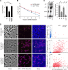
(p)ppGpp accumulation contributes to cell viability in swarmer cells during HipA2-induced growth arrest. (A) The indicated strains were grown to late exponential phase from approximately the same starting density. Cultures were then challenged with kanamycin (5 μg/ml) for 4 h. Results are shown as percent survival rate by comparison to untreated cells prior to the addition of antibiotic. Mean and SD from at least two independent experiments performed with three biological replicates (n = 6) are plotted. An asterisk indicates a significant difference (P < 0.05) by two-tailed Student’s t test. (B) The wild-type and ΔspoT strains harboring an extra copy of HA-HipA2 on the chromosome at the vanR locus were grown to the late exponential phase from approximately the same starting density. HipA2 expression was induced by the addition of 0.5 mM vanillate. Results show the percentage of survival at each indicated time point of vanillate treatment (log scale). Mean and SD from at least two independent experiments performed with three biological replicates (n = 6) are plotted. (C) Expression of HipA2 stimulates (p)ppGpp accumulation. Intracellular levels of (p)ppGpp extracted from the wild-type and ΔspoT strains expressing HipA2 toxin were detected by TLC. Mean and SD are plotted (n = 4). An asterisk indicates a significant difference (P < 0.05) by two-tailed Student’s t test in comparison with carbon starvation-treated wild-type cells. (D) SYTOX Deep Red staining reveals the persistence trait of swarmer cells in a (p)ppGpp-dependent manner. The indicated strains expressing DivJ-YFP were grown in PYE to the exponential phase. Cultures were supplemented with vanillate to induce HipA2 expression for 3 h and then challenged with kanamycin (5 μg/ml) for 4 h followed by SYTOX Deep Red staining according to manufacturer’s instructions. The arbitrary fluorescent intensities of YFP and SYTOX Deep Red are plotted.
FIG 4
FIG 4
HipA2 triggers an imbalance of intracellular free tryptophan and glutamine by stimulating GlnA adenylylation. (A) Schematic representation of GlnA adenylylation and deadenylylation. GlnA deadenylylation can be achieved by both hydrolysis (black arrow) and phosphorolysis (red arrow). (B) Stimulation of GlnA adenylylation by HipA2 analyzed by immunoprecipitation. Cells expressing HA-GlnA controlled by its native promoter and HipA2 controlled by the inducible xylose promoter were grown in PYE + glucose to exponential phase. Cells were collected, washed, and resuspended in PYE + xylose or PYE + glucose for induction or repression of HipA2 expression, respectively. Samples were collected at each indicated time point for immunoprecipitation using anti-HA magnetic beads and analyzed by SDS-PAGE and immunoprecipitation with either anti-AMP or anti-HA antibody. (C) Quantification of intracellular levels of free tryptophan and glutamine upon HipA2 activation. Strains harboring a chromosomal copy of Pxyl-hipA2 or Pxyl-hipA2D308Q (kinase-dead mutant) were grown in PYE + glucose to the exponential phase. Cells were collected, washed, and resuspended in PYE + xylose for HipA2 induction. Samples were collected at each indicated time point for free amino acid quantification, as described in Materials and Methods. (D) Comparison of GlnA adenylylation levels upon exposure to exogenous amino acids. Wild-type Caulobacter crescentus was grown in M2G medium to the exponential phase, and the culture was divided into three equal aliquots. The indicated amino acids were added to each aliquot, and cells were collected each hour for a period of 3 h. Lysates were prepared from each sample and analyzed by SDS-PAGE and immunoblotting with anti-AMP antibody. Due to the limited number of proteins that are adenylylated in Caulobacter (indicated by asterisks), we were able to compare levels of GlnA adenylylation in response to amino acid treatment.
FIG 5
FIG 5
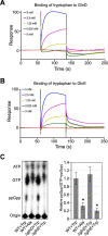
Tryptophan directly binds GlnD and GlnE to trigger (p)ppGpp accumulation. (A and B) Surface plasmon resonance (SPR) experiments show that tryptophan binds GlnD and GlnE. Purified GlnD or GlnE was tethered to the SPR chip and exposed to increasing concentrations of tryptophan. The binding period is displayed as response units plotted as function of time, followed by buffer injection to remove free tryptophan. Increasing concentrations of tryptophan used in the SPR experiments are shown. (C) Tryptophan stimulates (p)ppGpp accumulation. (Left) Intracellular levels of (p)ppGpp were detected by TLC. Alanine serves as a negative control. (Right) Relative concentrations of ppGpp as a function of tryptophan availability are shown. Mean and SD are plotted (n = 4). An asterisk indicates a significant difference (P < 0.05) by two-tailed Student’s t test in comparison with tryptophan-treated wild-type cells.
FIG 6
FIG 6
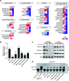
Caulobacter utilizes the stringent response to cope with multiple stresses by activation of HipA2 toxin. (A) Screening of environmental cues required for toxin activation in wild-type Caulobacter crescentus. Cultures were exposed to 69 different stress conditions and tested for their ability to induce upregulated transcription of hipB genes by qRT-PCR. The log-fold change [Log(FC)] of hipB1, hipB2, and hipB3 transcription in strains challenged by those stresses was plotted in a heat map. The category of stress conditions used for this study is indicated, and specific conditions, agents, and antibiotics are listed in Table S2. (B) Survival rates of the ΔhipBA2 mutant relative to the wild-type strain. Cells were grown to the exponential phase from approximately the same starting density. Cultures were then challenged with indicated stresses for 30 min (see Table S2 for detailed information) followed by the addition of kanamycin (5 μg/ml) for another 4 h. Results are shown as percent survival rate by comparison to untreated cells prior to the addition of the antibiotic. Mean and SD from one batch performed with three biological replicates (n = 6) are plotted. An asterisk indicates a significant difference (P < 0.05) by two-tailed Student’s t test. (C) The HipB2 protein is degraded by the Lon protease under conditions of exposure to heat shock, low pH, and carbon starvation. Wild-type (WT) and Δlon strains harboring a hipBA2 operon (expressing 6×His-hipB and native hipA) on a plasmid were grown in M2G to exponential phase. Cells were then suspended in the medium conferring the indicated stresses with an additional supplement of chloramphenicol (200 μg/ml) to shut off protein synthesis. Samples were collected every 20 min, and protein levels were monitored by immunoblot assays using anti-His antibody. 6×His-hipB was stable in Δlon cells. (D) In vivo phosphorylation of TrpS under the challenge of various stresses. Cells expressing TrpS-HA were treated with the indicated stresses for 3 h. Whole-cell lysates were analyzed by Phos-tag SDS-PAGE and immunoblotting with anti-HA antibody. Samples containing an empty vector or expressing HipA2 served as negative and positive controls, respectively.
FIG 7
FIG 7
Model of HipA2-induced amino acid imbalance that activates bacterial stringent response by stimulating GlnA (GS) adenylylation. Multiple stresses trigger the liberation of HipA2 toxin by activating Lon protease, which in turn mediates the proteolysis of antitoxin HipB2. The released HipA2 toxin deactivates tryptophanyl-tRNA synthetase TrpS and inhibits aminoacylation of tRNATrp by phosphorylation, yielding uncharged tRNATrp and free tryptophan repletion. The loading of uncharged tRNATrp at empty ribosomal A sites results in translation termination by stalled ribosomes. The high level of intracellular tryptophan triggers allosteric regulation of GlnD and GlnE by direct binding and thus stimulates GlnA adenylylation. GlnA-AMP is inactive and no longer catalyzes glutamine synthesis, leading to the accumulation of (p)ppGpp upon glutamine deprivation in a PTSNtr-dependent manner. HipA2-mediated toxicity enables Caulobacter to utilize the stringent response to cope with multiple stress conditions.

Similar articles

Cited by

References

    1. Gollan B, Grabe G, Michaux C, Helaine S. 2019. Bacterial persisters and infection: past, present, and progressing. Annu Rev Microbiol 73:359–385. doi:10.1146/annurev-micro-020518-115650. - DOI - PubMed
    1. Fisher RA, Gollan B, Helaine S. 2017. Persistent bacterial infections and persister cells. Nat Rev Microbiol 15:453–464. doi:10.1038/nrmicro.2017.42. - DOI - PubMed
    1. Kussell E, Kishony R, Balaban NQ, Leibler S. 2005. Bacterial persistence: a model of survival in changing environments. Genetics 169:1807–1814. doi:10.1534/genetics.104.035352. - DOI - PMC - PubMed
    1. Harms A, Maisonneuve E, Gerdes K. 2016. Mechanisms of bacterial persistence during stress and antibiotic exposure. Science 354:aaf4268. doi:10.1126/science.aaf4268. - DOI - PubMed
    1. Lewis K. 2010. Persister cells. Annu Rev Microbiol 64:357–372. doi:10.1146/annurev.micro.112408.134306. - DOI - PubMed

Publication types

LinkOut - more resources