Case Reports
. 2021 Mar-Apr;35(2):913-918.
doi: 10.21873/invivo.12331.

Laparoscopic Distal Gastrectomy for Synchronous Gastric Cancer and Gastrointestinal Stromal Tumor With Situs Inversus Totalis

Affiliations
Case Reports

Laparoscopic Distal Gastrectomy for Synchronous Gastric Cancer and Gastrointestinal Stromal Tumor With Situs Inversus Totalis

Tsutomu Namikawa et al. In Vivo. 2021 Mar-Apr.

Abstract

Background: Situs inversus totalis (SIT) is a rare congenital condition in which the thoracic and abdominal organs are inverted like a mirror image.

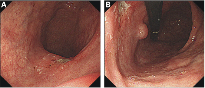
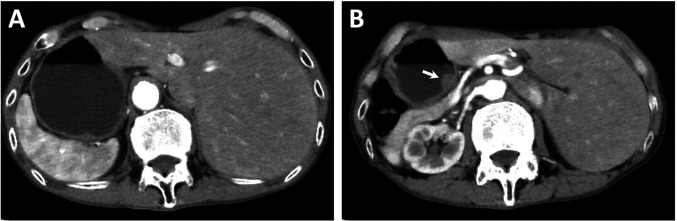
Case report: We present a case of synchronous gastric cancer and gastrointestinal stromal tumor (GIST) associated with SIT in a 74-year-old man who was admitted to our department to treat gastric cancer. Esophagogastroduodenoscopy revealed a depressed lesion and a submucosal tumor (SMT) in the middle-third of the stomach. Abdominal contrast-enhanced computed tomography revealed complete inversion of the internal organs, and the common hepatic artery branched from the superior mesenteric artery. The patient underwent laparoscopic distal gastrectomy with regional lymph node dissection and Billroth I reconstruction. The macroscopic observation of the resected specimen revealed a depressed lesion measuring 2.0×1.5 cm in diameter and an SMT measuring 2.2×1.8 cm.

Conclusion: Careful preoperative anatomic evaluation is important in SIT because the situs anomalies may be accompanied by major vascular anomalies.

Keywords: Situs inversus; gastric cancer; gastrointestinal stromal tumor; laparoscopic distal gastrectomy; three-dimensional computed tomography.


Conflict of interest statement

The Authors have no conflicts of interest to declare regarding this study.

Figures

Figure 1
Figure 1. Esophagogastroduodenoscopy shows a superficial depressing-type tumor and a submucosal tumor. A shallow ulcer (A and B) and a submucosal tumor (B) are observed on the posterior wall of the middle-third of the stomach.
Figure 2
Figure 2. Abdominal contrast-enhanced computed tomography shows situs inversus totalis. Complete transposition of the abdominal viscera (A and B), with a mass protruding into the gastric lumen (B, arrow).
Figure 3
Figure 3. Three-dimensional reconstruction image of computed tomography angiography (3DCTA) and double-contrast barium imaging. 3DCTA shows complete transposition of vessels: the common hepatic artery is noted to branch from the superior mesenteric artery (A, arrow). Doublecontrast barium imaging shows a lesion of the deformed wall, with gastric fold concentration, in the posterior wall (B, white arrowhead) and an intragastric protruding lesion in the lesser curvature of the middle-third of the stomach (B, black arrowhead).
Figure 4
Figure 4. Intraoperative findings during laparoscopic distal gastrectomy. The stomach and spleen are located on the right side of the abdomen (A and B). After lymph node dissection, the splenic artery is noted on the right side of the patient (C, arrow), stumps of the left gastric artery and vein are shown. The splenic vein is located on the upper edge of the pancreas because the common hepatic artery branched from the superior mesenteric artery (D, arrowhead).
Figure 5
Figure 5. Gross examination of the surgically resected specimen. The resected specimen reveals a slightly depressed lesion measuring 21×13 mm (arrowheads) and a submucosal tumor measuring 22×18 mm (arrow)

Similar articles

Cited by

References

    1. Kigasawa Y, Takeuchi H, Kawakubo H, Fukuda K, Nakamura R, Takahashi T, Wada N, Kitagawa Y. Laparoscopy-assisted distal gastrectomy in a case of gastric cancer with situs inversus totalis: a case report. Asian J Endosc Surg. 2017;10(1):47–50. doi: 10.1111/ases.12326. - DOI - PubMed
    1. Namikawa T, Tsuda S, Fujisawa K, Iwabu J, Uemura S, Tsujii S, Maeda H, Kitagawa H, Kobayashi M, Hanazaki K. Superficial spreading-type gastric cancer with situs inversus totalis. In Vivo. 2018;32(3):685–689. doi: 10.21873/invivo.11294. - DOI - PMC - PubMed
    1. Fletcher CD, Berman JJ, Corless C, Gorstein F, Lasota J, Longley BJ, Miettinen M, O’Leary TJ, Remotti H, Rubin BP, Shmookler B, Sobin LH, Weiss SW. Diagnosis of gastrointestinal stromal tumors: A consensus approach. Hum Pathol. 2002;33(5):459–465. doi: 10.1053/hupa.2002.123545. - DOI - PubMed
    1. Yamaguchi S, Orita H, Yamaoka T, Mii S, Sakata H, Hashizume M. Laparoscope-assisted distal gastrectomy for early gastric cancer in a 76-year-old man with situs inversus totalis. Surg Endosc. 2003;17(2):352–353. doi: 10.1007/s00464-002-4504-y. - DOI - PubMed
    1. Futawatari N, Kikuchi S, Moriya H, Katada N, Sakuramoto S, Watanabe M. Laparoscopy-assisted distal gastrectomy for early gastric cancer with complete situs inversus: report of a case. Surg Today. 2010;40(1):64–67. doi: 10.1007/s00595-009-4007-8. - DOI - PubMed

Publication types

LinkOut - more resources