

Pelvic Insufficiency Fractures in Cervical Cancer After Radiation Therapy: A Meta-Analysis and Review
- PMID: 33622908
- PMCID: PMC8045108
- DOI: 10.21873/invivo.12356
Pelvic Insufficiency Fractures in Cervical Cancer After Radiation Therapy: A Meta-Analysis and Review
Abstract
Aim: The aim of the study was to estimate the prevalence of pelvic insufficiency fractures (PIFs) after radiation therapy (RT) in patients with cervical cancer.
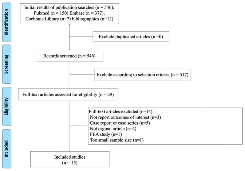
Patients and methods: A total of 3,633 patients from 15 cohort studies were included. Proportion meta-analysis was performed to estimate prevalence and subgroup analysis was performed according to imaging modalities for diagnosis of PIF. For continuous variables (age and length of follow-up), meta-regression analysis was performed.
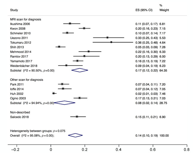
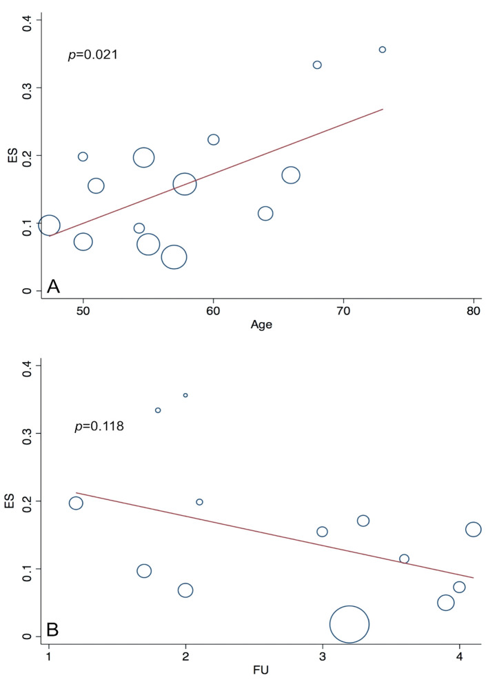
Results: Pooled prevalence estimate of PIF was 14% (95% CI=10-19). Incidence of PIF was higher in studies that used MRI as a diagnostic tool (17%, 95% CI=12-22) than non-MRI (8%, 95% CI=2-14). In meta-regression, we found a significant association of prevalence of PIF with age (p=0.021) but not with length of follow-up (p=0.118).
Conclusion: PIF after RT in patients with cervical cancer is not rare. Physicians need to pay attention to PIFs, especially in patients with high-risk factors for osteoporotic fracture.
Keywords: Cervical cancer; insufficiency fracture; osteoporosis; survivoral care.
Copyright© 2021, International Institute of Anticancer Research (Dr. George J. Delinasios), All rights reserved.
Conflict of interest statement
The Authors declare no conflicts of interest.
Figures
Similar articles
-
Risk Factors for Pelvic Insufficiency Fractures in Locally Advanced Cervical Cancer Following Intensity Modulated Radiation Therapy.Int J Radiat Oncol Biol Phys. 2017 Apr 1;97(5):1032-1039. doi: 10.1016/j.ijrobp.2017.01.026. Epub 2017 Mar 15. Int J Radiat Oncol Biol Phys. 2017. PMID: 28332986
-
Pelvic fractures after definitive and postoperative radiotherapy for cervical cancer: A retrospective analysis of risk factors.Gynecol Oncol. 2017 Dec;147(3):585-588. doi: 10.1016/j.ygyno.2017.09.035. Epub 2017 Oct 18. Gynecol Oncol. 2017. PMID: 29055558
-
Insights into pelvic insufficiency fracture following pelvic radiotherapy for cervical cancer: a comparative review.BMC Womens Health. 2024 May 23;24(1):306. doi: 10.1186/s12905-024-03099-8. BMC Womens Health. 2024. PMID: 38783273 Free PMC article. Review.
-
Radiation-Induced Insufficiency Fractures After Pelvic Irradiation for Gynecologic Malignancies: A Systematic Review.Int J Radiat Oncol Biol Phys. 2020 Nov 1;108(3):620-634. doi: 10.1016/j.ijrobp.2020.05.013. Epub 2020 May 19. Int J Radiat Oncol Biol Phys. 2020. PMID: 32442476
-
Pelvic insufficiency fractures in patients with cervical and endometrial cancer treated with postoperative pelvic radiation.Gynecol Oncol. 2013 Mar;128(3):540-3. doi: 10.1016/j.ygyno.2012.12.021. Epub 2012 Dec 20. Gynecol Oncol. 2013. PMID: 23262211
Cited by
-
Pelvic Insufficiency Fractures and Bone Pain after Radiation Therapy for Anal Cancer: Relation to Pelvic Bone Dose-Volume Parameters.Adv Radiat Oncol. 2022 Oct 20;8(1):101110. doi: 10.1016/j.adro.2022.101110. eCollection 2023 Jan-Feb. Adv Radiat Oncol. 2022. PMID: 36483064 Free PMC article.
-
Detection of vertebral insufficiency fractures on 18F-FDG PET/CT following radiation therapy for oesophageal cancer.Radiol Case Rep. 2025 Jun 27;20(9):4669-4673. doi: 10.1016/j.radcr.2025.06.022. eCollection 2025 Sep. Radiol Case Rep. 2025. PMID: 40677874 Free PMC article.
-
Superiority of MRI for Evaluation of Sacral Insufficiency Fracture.J Clin Med. 2022 Aug 24;11(17):4968. doi: 10.3390/jcm11174968. J Clin Med. 2022. PMID: 36078896 Free PMC article.
-
Therapy-induced bone changes in oncology imaging with 18F-sodium fluoride (NaF) PET-CT.Ann Nucl Med. 2022 Apr;36(4):329-339. doi: 10.1007/s12149-022-01730-y. Epub 2022 Feb 26. Ann Nucl Med. 2022. PMID: 35218508 Review.
-
Relations Between Bone Quantity, Microarchitecture, and Collagen Cross-links on Mechanics Following In Vivo Irradiation in Mice.JBMR Plus. 2021 Sep 26;5(11):e10545. doi: 10.1002/jbm4.10545. eCollection 2021 Nov. JBMR Plus. 2021. PMID: 34761148 Free PMC article.
References
-
- Cancer fact sheets: Cervix uteri. Geneva, World Health Organization: Internationanl Agency for Research on Cancer. 2020. Available at: https://gco.iarc.fr/today/data/factsheets/cancers/23-Cervix-uteri-fact-s... [Last accessed on 1st January 2021]
-
- Klaitong C, Meannuch E, Kaewbunperm U, Klaiphibule P, Sinthusek T. Radiotherapy at pelvis region in menopausal cervix cancer induce osteopenia/osteoporosis. Radiother Oncol. 2018;127:S819–S820. doi: 10.1016/S0167-8140(18)31821-8. - DOI
Publication types
MeSH terms
LinkOut - more resources
Full Text Sources
Other Literature Sources
Medical