. 2021 Feb 16:15:645-650.
doi: 10.2147/OPTH.S294966. eCollection 2021.

Femtosecond Laser-Assisted Big-Bubble Deep Anterior Lamellar Keratoplasty

Affiliations

Femtosecond Laser-Assisted Big-Bubble Deep Anterior Lamellar Keratoplasty

Emilio Pedrotti et al. Clin Ophthalmol. .

Abstract

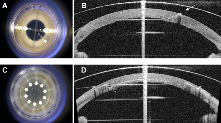
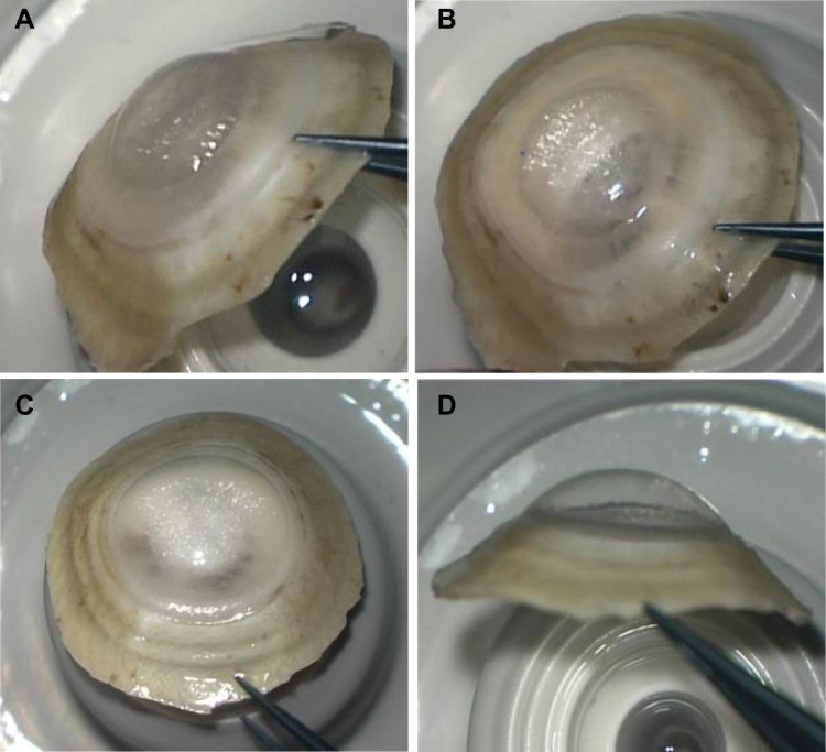
Purpose: To determine whether type 1 big-bubble (BB) formation is influenced by the sequence of incisions created with the Victus femtosecond laser (FSL) enabled with software version 3.4 (SV 3.4) during deep anterior lamellar keratoplasty (DALK).

Materials and methods: Consecutive FSL-assisted DALK BB procedures were performed on 20 human donor corneas: 10 shaped by tunnel incision followed by lamellar incision (tunnel-lamellar group, TL) and 10 in the reverse order (lamellar-tunnel group, LT). The BB type was assessed by evaluating dynamic air movement during air inflation; bubble diameter and floor thickness were measured by anterior segment optical coherence tomography.

Results: Overall, a type 1 BB formed in 85% of eyes: 100% in the TL group and 70% in the LT group. In the LT group, a type 2 BB formed in 2 corneas and one cornea was perforated during cannula insertion. Type 1 BB was achieved after one attempt in 90% of eyes in the TL group and in 57% in the LT group.

Conclusion: Shaping the tunnel before rather than after lamellar incision may be more effective for obtaining a type 1 BB by air injection.

Keywords: big bubble; deep anterior lamellar keratoplasty; femtosecond laser; intrastromal incision; lamellar incision.


Conflict of interest statement

The author reports no conflicts of interest. The study was supported by Bausch & Lomb.

Figures

Figure 1
Figure 1
Surgical microscopy and integrated optical coherence tomography images of tunnel and mushroom FSL incisions. Frontal view of the tunnel incision in the paracentral position at 130° (A, arrowhead); plane 1 is angulated while plane 2 lies parallel to the corneal surface (B, arrow); the cut of the internal layers is clear from the frontal view (C); anterior, middle, and posterior view of the lamellar cut following mushroom configuration (D).
Figure 2
Figure 2
Appearance of type 1 and type 2 BB after eversion of the cornea. Type 1 BB, frontal (A) and lateral view (B); type 2 BB, frontal (C) and lateral view (D) showing a smaller diameter type 1 compared to type 2 bubble.

Similar articles

Cited by

References

    1. Ple-Plakon PA, Shtein RM. Trends in corneal transplantation: indications and techniques. Curr Opin Ophthalmol. 2014;25:300–305. doi:10.1097/ICU.0000000000000080 - DOI - PubMed
    1. Reinhart WJ, Musch DC, Jacobs DS, et al. Deep anterior lamellar keratoplasty as an alternative to penetrating keratoplasty. Ophthalmology. 2011;118:209–218. doi:10.1016/j.ophtha.2010.11.002 - DOI - PubMed
    1. Anwar M, Teichmann KD. Big-bubble technique to bare Descemet’s membrane in anterior lamellar keratoplasty. J Cataract Refract Surg. 2002;28:398–403. doi:10.1016/S0886-3350(01)01181-6 - DOI - PubMed
    1. Dua HS, Faraj LA, Kenawy MB, et al. Dynamics of big bubble formation in deep anterior lamellar keratoplasty by the big bubble technique: in vitro studies. Acta Ophthalmol. 2018;96:69–76. doi:10.1111/aos.13460 - DOI - PubMed
    1. Myerscough J, Friehmann A, Bovone C, Mimouni M, Busin M. Management of type 2 bubble formed during big-bubble deep anterior lamellar keratoplasty. Cornea. 2019;38(6):E20. doi:10.1097/ICO.0000000000001940 - DOI - PubMed

LinkOut - more resources