. 2021 Feb 15:14:671-682.
doi: 10.2147/DMSO.S288338. eCollection 2021.

Overexpression of miR125b Promotes Osteoporosis Through miR-125b-TRAF6 Pathway in Postmenopausal Ovariectomized Rats

Affiliations

Overexpression of miR125b Promotes Osteoporosis Through miR-125b-TRAF6 Pathway in Postmenopausal Ovariectomized Rats

Gang Wang et al. Diabetes Metab Syndr Obes. .

Abstract

Background: Postmenopausal osteoporosis is one of the most common types of osteoporosis that women suffer from. Studies involving molecular mechanisms for designing better therapeutic strategies for postmenopausal osteoporosis are still rare. The present study investigates the role of miR-125b in postmenopausal osteoporosis.

Methods: Microarray analysis was done to screen the gene database. Tissue samples of postmenopausal women were collected to study the miRNA profiles. MC3T3-E1 cells were used and were submitted for transfection. CCK-8 assay was done to check the viability of cells, whereas toxicity was done by lactate dehydrogenase assay kit. TargetScan was done to target genes of miR-125b followed by confirmation by Luciferase reporter assay. For animal studies a rat model of ovariectomized rats was created. Bone mineral density and biomechanics were measured by densitometer. The mRNA levels were assessed by qRT-PCR and proteins by Western blot assay.

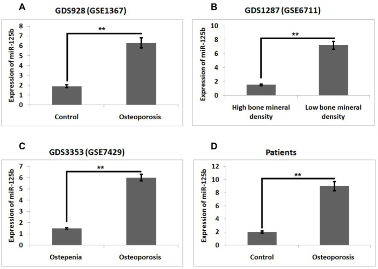
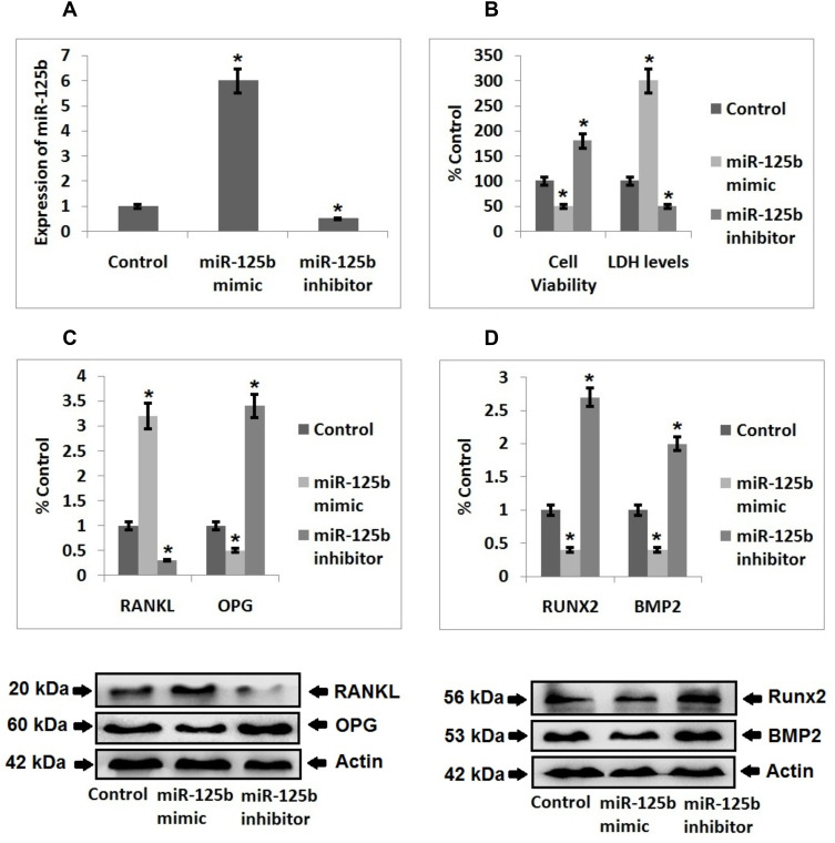
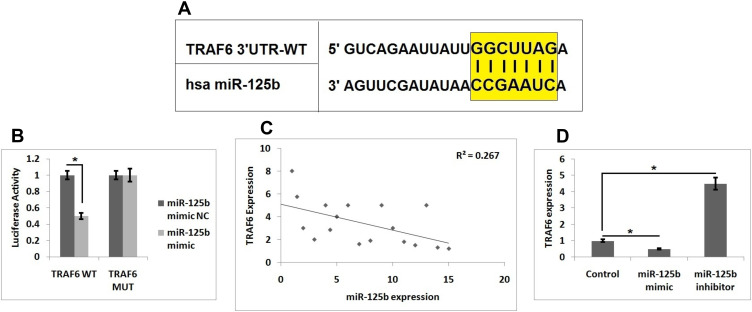
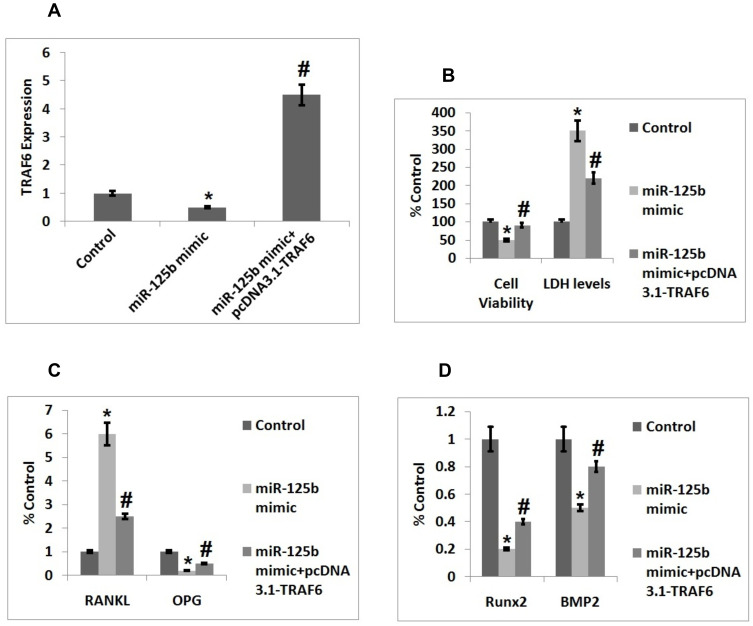
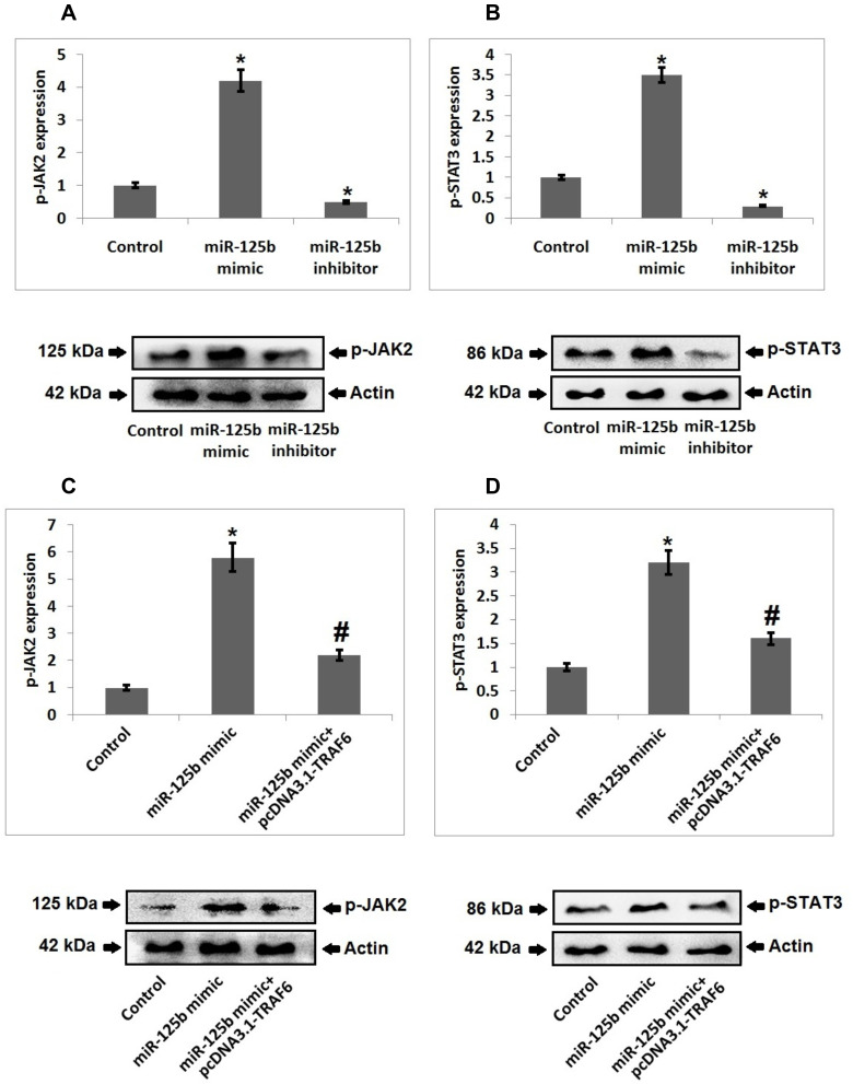
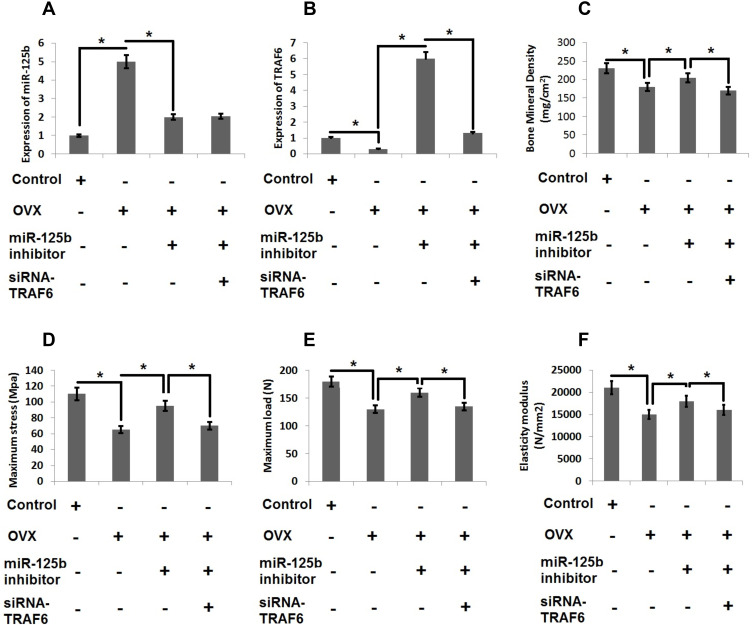
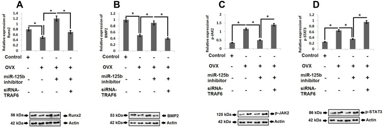
Results: miR-125b was over-expressed in human osteoporosis samples. In vitro studies suggested that miR-125b suppressed the cell viability and promoted release of LDH, it also enhanced the RANKL/OPG ratio and suppressed levels of BMP2 and Runx2. Bioinformatics identified TRAF6 as a potential target of miR-125b, further confirmed by luciferase assay, also miR-125b negatively regulated the levels of TRAF6 gene in osteoporosis bones involving the JAK2/STAT3 cascade. In the rat model, miR-125b decreased the bone mineral density and biomechanical parameters in bones by altering the TRAF6 gene involving the JAK2/STAT3 pathway.

Conclusion: The outcomes suggested that miR-125b was responsible for the development of postmenopausal osteoporosis and promoted its progression by the TRAF6 gene via the JAK2/STAT3 pathway.

Keywords: JAK2/STAT3 pathway; TRAF6; miR‐125b; osteoporosis.


Conflict of interest statement

The authors declare no conflicts of interest for this work.

Figures

Figure 1
Figure 1
miR‐125b is overexpressed in postmenopausal osteoporosis. Levels of miR-125b in (A) GDS928 (GSE1367) dataset, (B) GDS1287 (GSE6711), (C) DS3353 (GSE7429) and (D) in clinical samples. The results are mean ± SD, **P<0.01 compared to control.
Figure 2
Figure 2
miR‐125b induced osteoporosis in MC3T3-E1 cells. (A) Quantitative results of qRT-PCR showing transfection efficiency. (B) Quantitative results showing viability of cells and levels of LDH after 48 hours post transfection. (C) Western blot analysis for expression of RANKL, OPG in the MC3T3-E1 cells and quantitative results showing the expression of proteins. (D) Western blot analysis for expression of Runx2 and BMP2 in the cells and quantitative results showing expression of proteins. The results are mean ± SD, *P<0.05 compared to control.
Figure 3
Figure 3
TRAF6 is the target gene of miR-125b. (A) The 3ʹUTR region of TRAF6 has potential binding site for miR-125b. (B) Results of Luciferase assay as evaluated in MC3T3-E1 cells. (C) Results of Pearson correlation analysis for levels of miR-125b levels and TRAF6 in osteoporosis clinical samples. (D) Levels of TRAF6 in MC3T3-E1 cells. The results are mean ± SD. *P<0.05 compared to control.
Figure 4
Figure 4
Upregulation of TRAF6 inverts the alterations of cell function mediated by miR-125b. (A) Quantitative results of qRT-PCR showing transfection efficacy of TRAF6. (B) Quantitative results showing viability of cells and levels of LDH after 48 post transfection. (C) Quantitative results showing expression of proteins RANKL and OPG in cells. (D) Quantitative results showing expression of Runx2 and BMP2 in cells. The results are mean ± SD. *P<0.05 compared to control, #P<0.05 compared to miR-125b mimic.
Figure 5
Figure 5
JAK2/STAT3 cascade is responsible for miR-125b-TRAF6 mediated osteoporosis. (A) Western blot analysis and quantitative results showing expression of p-JAK2 in miR-125b mimic/inhibitor treated cells. (B) Western blot analysis and quantitative results showing expression of p-STAT3 in miR-125b mimic/inhibitor treated cells. (C) Western blot analysis and quantitative results showing expression of p-JAK2 in miR-125b mimic treated cells and mimic+ pcDNA3.1-TRAF6 transfected cells. (D) Western blot analysis and quantitative results showing expression of p-STAT3 in miR-125b mimic + pcDNA3.1-TRAF6 transfected cells. The results are mean ± SD. *P<0.05 compared to miR-125b mimic treated group, #<0.05 compared to control.
Figure 6
Figure 6
miR-125b-TRAF6 pathway regulates the progression of osteoporosis in OVX rats. (A) miR-125b levels were evaluated by qRT-PCR analysis. (B) mRNA levels of TRAF6 by qRT-PCR analysis. (C) Results of bone mineral density of isolated femurs. (D) Maximum bending stress of femur bones. (E) The maximum load bearing capacity of femur bones. (F) Results of elasticity modulus of femur bones. The results are mean ± SD. *P<0.05 compared with adjacent groups.
Figure 7
Figure 7
JAK2/STAT3 cascade is the downstream target of miR-125b-TRAF6 pathway in OVX rats. Western blot analysis and quantitative analysis for expression Runx2 (A), BMP-2 (B), p-JAK2 (C) and pSTAT3 (D) in OVX rats. The results are mean ± SD. *P<0.05 compared to adjacent groups.

Similar articles

Cited by

References

    1. Compston JE, Mcclung MR, Leslie WD. Osteoporosis. Lancet. 2019;393:364–376. doi:10.1016/S0140-6736(18)32112-3 - DOI - PubMed
    1. Xu X, Jia X, Mo L, et al. Intestinal microbiota: a potential target for the treatment of postmenopausal osteoporosis. Bone Res. 2017;5:17046. - PMC - PubMed
    1. Chen X, Zhang S, Chen X, et al. Emodin promotes the osteogenesis of MC3T3‐E1 cells via BMP‐9/Smad pathway and exerts a preventive effect in ovariectomized rats. Acta Biochimica et Biophysica Sinica. 2017;49:867–878. - PubMed
    1. Eastell R, O’Neill T, Hofbauer L, et al. Postmenopausal osteoporosis. Nat Rev Dis Primers. 2016;2:1–16. - PubMed
    1. Black DM, Rosen CJ. Postmenopausal osteoporosis. N Engl J Med. 2016;374:254‐262. - PubMed

LinkOut - more resources