

Role of 18F-FDG positron emission tomography in carotid atherosclerotic plaque imaging: A systematic review
- PMID: 33623500
- PMCID: PMC7875029
- DOI: 10.4103/wjnm.WJNM_26_20
Role of 18F-FDG positron emission tomography in carotid atherosclerotic plaque imaging: A systematic review
Abstract
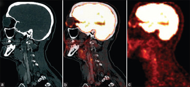
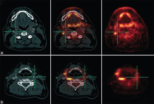
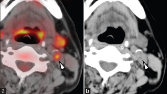
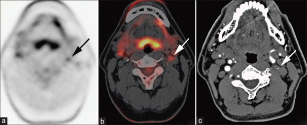
Stroke and other thromboembolic events in the brain are often due to carotid artery atherosclerosis, and atherosclerotic plaques with inflammation are considered particularly vulnerable, with an increased risk of becoming symptomatic. Positron emission tomography (PET) with 2-deoxy-2-[Fluorine-18] fluoro-D-glucose (18F-FDG) provides valuable metabolic information regarding arteriosclerotic lesions and may be applied for the detection of vulnerable plaque. At present, however, patients are selected for carotid surgical intervention on the basis of the degree of stenosis alone, and not the vulnerability or inflammation of the lesion. During the past decade, research using PET with the glucose analog tracer 18F-fluor-deoxy-glucose, has been implemented for identifying increased tracer uptake in symptomatic carotid plaques, and tracer uptake has been shown to correlate with plaque inflammation and vulnerability. These findings imply that 18F-FDG PET might hold the promise for a new and better diagnostic test to identify patients eligible for carotid endarterectomy. The rationale for developing diagnostic tests based on molecular imaging with 18F-FDG PET, as well as methods for simple clinical PET approaches, are discussed. This is a systematic review, following Preferred Reporting Items for Systematic Reviews guidelines, which interrogated the PUBMED database from January 2001 to November 2019. The search combined the terms, "atherosclerosis," "inflammation," "FDG," and "plaque imaging." The search criteria included all types of studies, with a primary outcome of the degree of arterial vascular inflammation determined by 18F-FDG uptake. This review examines the role of 18F-FDG PET imaging in the characterization of atherosclerotic plaques.
Keywords: 18F-FDG; atherosclerosis; carotid artery; carotid endarterectomy; plaque imaging; vulnerable plaque.
Copyright: © 2020 World Journal of Nuclear Medicine.
Conflict of interest statement
There are no conflicts of interest.
Figures
Similar articles
-
Carotid plaque inflammation assessed with (18)F-FDG PET/CT is higher in symptomatic compared with asymptomatic patients.Int J Stroke. 2015 Jul;10(5):730-6. doi: 10.1111/ijs.12430. Epub 2015 Jan 15. Int J Stroke. 2015. PMID: 25588553
-
High-risk plaque features can be detected in non-stenotic carotid plaques of patients with ischaemic stroke classified as cryptogenic using combined (18)F-FDG PET/MR imaging.Eur J Nucl Med Mol Imaging. 2016 Feb;43(2):270-279. doi: 10.1007/s00259-015-3201-8. Epub 2015 Oct 3. Eur J Nucl Med Mol Imaging. 2016. PMID: 26433367
-
Cohort profile: BIOVASC-late, a prospective multicentred study of imaging and blood biomarkers of carotid plaque inflammation and risk of late vascular recurrence after non-severe stroke in Ireland.BMJ Open. 2020 Jul 19;10(7):e038607. doi: 10.1136/bmjopen-2020-038607. BMJ Open. 2020. PMID: 32690537 Free PMC article.
-
Carotid Plaque Positron Emission Tomography Imaging and Cerebral Ischemic Disease.Stroke. 2019 Aug;50(8):2072-2079. doi: 10.1161/STROKEAHA.118.023987. Epub 2019 Jul 5. Stroke. 2019. PMID: 31272325 Free PMC article.
-
Molecular imaging of carotid artery atherosclerosis with PET: a systematic review.Eur J Nucl Med Mol Imaging. 2020 Jul;47(8):2016-2025. doi: 10.1007/s00259-019-04622-y. Epub 2019 Nov 30. Eur J Nucl Med Mol Imaging. 2020. PMID: 31786626
Cited by
-
In vitro and pilot in vivo imaging of 18 kDa translocator protein (TSPO) in inflammatory vascular disease.EJNMMI Res. 2021 May 5;11(1):45. doi: 10.1186/s13550-021-00786-7. EJNMMI Res. 2021. PMID: 33950298 Free PMC article.
-
Diagnostics of atherosclerosis: Overview of the existing methods.Front Cardiovasc Med. 2023 May 9;10:1134097. doi: 10.3389/fcvm.2023.1134097. eCollection 2023. Front Cardiovasc Med. 2023. PMID: 37229223 Free PMC article. Review.
-
Effect of statins on arterial wall inflammation as assessed by 18F-FDG PET CT: an updated systematic review and meta-analysis.J Inflamm (Lond). 2024 Dec 18;21(1):52. doi: 10.1186/s12950-024-00421-x. J Inflamm (Lond). 2024. PMID: 39696570 Free PMC article.
-
Preclinical models of radiation-induced cardiac toxicity: Potential mechanisms and biomarkers.Front Oncol. 2022 Oct 12;12:920867. doi: 10.3389/fonc.2022.920867. eCollection 2022. Front Oncol. 2022. PMID: 36313656 Free PMC article. Review.
References
Publication types
LinkOut - more resources
Full Text Sources
Miscellaneous