

Detection of muscle metastases on 18F-fluorodeoxyglucose positron emission tomography/computed tomography scan in 13 cases
- PMID: 33623509
- PMCID: PMC7875027
- DOI: 10.4103/wjnm.WJNM_61_19
Detection of muscle metastases on 18F-fluorodeoxyglucose positron emission tomography/computed tomography scan in 13 cases
Abstract
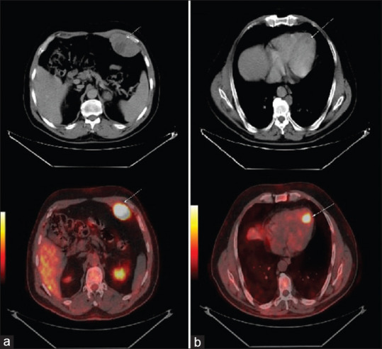
Muscular metastases (MMs) form an infrequent entity, and their physiopathology is still not well-defined. In this study, we estimated the incidence of MMs that were detected by 18F-fluorodeoxyglucose positron emission tomography/computed tomography and also specified their metabolic characteristics. This study includes 13 patients with MMs from a remotely located primary tumor. The results of this study showed an incidence of MMs at about 1%, with the most frequently involved muscles being iliopsoas and paraspinal. Lung cancer seems to be the most common tumor that causes MMs. Furthermore, these MMs vary in size and physiological uptake; they seem to be out of the ordinary and easily detected. They are often associated with other extra muscular locations and frequently involve the trunk muscles. Their detection in the course of the evolution of a specific neoplasia testifies to their aggressiveness and portends an unfavorable prognosis. The data in our series confirm that in the literature regarding the underlying primary tumors and anatomical sites involved by MMs.
Keywords: 18F-fluorodeoxyglucose; iliopsoas; muscular metastases; neoplasia; paraspinal; physiological uptakes; positron emission tomography/computed tomography.
Copyright: © 2020 World Journal of Nuclear Medicine.
Conflict of interest statement
There are no conflicts of interest.
Figures


References
-
- So Y, Yi JG, Song I, Lee WW, Chung HW, Park JH, et al. Detection of skeletal muscle metastasis: Torso FDG PET-CT versus contrast-enhanced chest or abdomen CT. Acta Radiol. 2015;56:860–6. - PubMed
-
- Haygood TM, Wong J, Lin JC, Li S, Matamoros A, Costelloe CM, et al. Skeletal muscle metastases: A three-part study of a not-so-rare entity. Skeletal Radiol. 2012;41:899–909. - PubMed
-
- Sudo A, Ogihara Y, Shiokawa Y, Fujinami S, Sekiguchi S. Intramuscular metastasis of carcinoma. Clin Orthop Relat Res. 1993;296:213–7. - PubMed
LinkOut - more resources
Full Text Sources