Review
. 2020 Oct 10;14(2):526-536.
doi: 10.1093/ckj/sfaa142. eCollection 2021 Feb.

Acute kidney injury pathology and pathophysiology: a retrospective review

Affiliations
Review

Acute kidney injury pathology and pathophysiology: a retrospective review

Joseph P Gaut et al. Clin Kidney J. .

Abstract

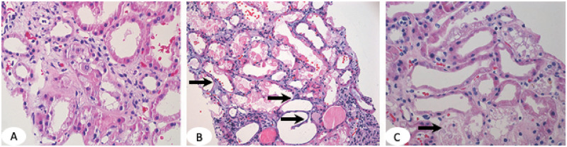
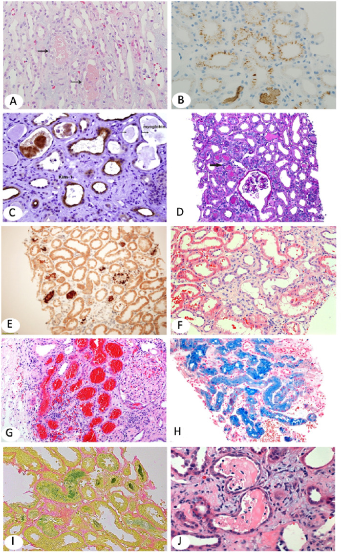
Acute kidney injury (AKI) is the clinical term used for decline or loss of renal function. It is associated with chronic kidney disease (CKD) and high morbidity and mortality. However, not all causes of AKI lead to severe consequences and some are reversible. The underlying pathology can be a guide for treatment and assessment of prognosis. The Kidney Disease: Improving Global Outcomes guidelines recommend that the cause of AKI should be identified if possible. Renal biopsy can distinguish specific AKI entities and assist in patient management. This review aims to show the pathology of AKI, including glomerular and tubular diseases.

Keywords: AKI; acute tubular necrosis; hemoglobinuria; multiple myeloma; pathology; pathophysiology; review; rhabdomyolysis.


Figures

FIGURE 1
FIGURE 1
(A) ATI in proximal tubules shows luminal dilatation, simplification of the lining epithelium and loss of epithelial cell nuclei in some cells and loss of the brush border. (B) ATN is defined by tubular epithelial cell necrosis (dark pink fragmented cytoplasm with no nuclei) and denudation of the basement membrane (arrows). (C) ATI and ATN in the same renal biopsy. Arrow points to necrotic tubules. Dilated tubules are lined by a thin epithelial layer with no brush border. H&E,×100.
FIGURE 2
FIGURE 2
(A) Myoglobin casts involve focal tubules and appear light pink on H&E (×100). Arrows point to myoglobin casts. (B) Myoglobin stains tubular casts brown and may also stain tubular epithelial brush border and/or cytoplasm in a punctuate pattern. Immunohistochemistry (IHC) ×100. (C) KIM-1, a marker for AKI, is overexpressed in injured and simplified (thin) tubular epithelium [same biopsy as in (B)]. KIM-1 IHC ×200. (D, E) The biopsy shows ATI with focal translucent tubular casts (arrow in D). Hemoglobin IHC highlights the tubular casts (E). Myoglobin stain was negative. The patient in (D–E), a 72-year-old Caucasian man with severe coronary artery disease, hypertension (HTN) and type 2 diabetes developed recurrent infection on his right foot, treated with intravenous piperacillin/tazobactam and developed chills and shortness of breath. He also had hematuria and severe peripheral hemolysis. CPK was normal; creatinine increased to 7 mg/dL with low C3 and C4. Clinical diagnoses included all comorbidities, but hemoglobin nephropathy was least expected. Hemoglobin IHC ×100. (F) Patient with IgA nephropathy who presented with hematuria and AKI. Renal biopsy shows tubular dilatation, simplification of the epithelium and multifocal luminal RBCs (H&E ×100). (G) Large patch of subcapsular proximal tubules packed with RBCs. Renal biopsy is from a 79-year-old white woman who presented with AKI on CKD. She has a histroy of atrial fibrillation on Coumadin. (I) Faucet stain marking bilirubin casts (×100). The patient was a 50-year-old Caucasian man with kidney transplant and AKI. Serum creatinine was 3.9 mg/dL and bilirubin and liver function tests were increased. (H) Marked tubular iron deposits with Prussian blue stain. The patient is a 60-year-old African American man who presented with AKI, macroscopic hematuria, hemolysis 1+ and increased reticulocytes. He had a history of mitral valve replacement, congestive heart failure and anemia. The differential diagnosis included cardiac valve defect, sickle cell disease and/or supratherapeutic international normalized ratio (H&E ×100). (JL) Diffuse ATI and typical multiple myeloma casts that appear as partially crumbled luminal protein deposits admixed with inflammatory cells.

References

    1. Brun CC, Munck O. Lesions of the kidney in acute renal failure following shock. Lancet 1957; 269: 603–607 - PubMed
    1. Smith HM. The Kidney: Structure and Function in Health and Disease. 1st edn. Oxford: Oxford University Press, 1951
    1. Bellomo R, Kellum JA, Ronco C. Defining acute renal failure: physiological principles. Intensive Care Med 2004; 30: 33–37 - PubMed
    1. Ronco C, Bellomo R, Kellum JA. Acute kidney injury. Lancet 2019; 394: 1949–1964 - PubMed
    1. Kidney Disease: Improving Global Outcomes Acute Kidney Injury Work Group. KDIGO Clinical Practice Guideline for Acute Kidney Injury. Kidney Int Suppl 2012; 2: 1–138