

Ulnar Nerve and Ulnar Artery Injury Caused by Comminuted Distal Radius Fracture
- PMID: 33623761
- PMCID: PMC7885659
- DOI: 10.13107/jocr.2020.v10.i04.1786
Ulnar Nerve and Ulnar Artery Injury Caused by Comminuted Distal Radius Fracture
Abstract
Introduction: Distal radius fractures are one of the most frequent traumas encountered in daily orthopedic practice. With this case report, we would like to emphasize the significance of an unexpected associated ulnar nerve and artery injury with distal radius fracture to physicians.
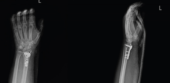
Case report: A 56-year-old male patient was evaluated in the emergency room after a motorcycle accident. The left wrist had a deformity and swelling, and about 3 × 1.5 cm of superficial skin abrasion was found in the volar surface of the wrist. It was noted that distal pulses were palpable, no neurological damage was found except hypoesthesia in the 5th finger. Radiologic examination revealed that the right shoulder was dislocated, and there was a displaced comminuted distal radius fracture in the left wrist with a non-displaced fracture of the ulnar styloid. The fracture was treated with open reduction and internal fixation using volar anatomic plate through the volar approach. After the surgery, pre-operative numbness did not resolve and opposing that expected; it increases with associated pain on the ulnar nerve innervated area within 30 days. The electromyographic analysis revealed severe partial ulnar nerve injury. The surgical exploration of the nerve was decided. The ulnar nerve was found to be trapped in scar tissue, and intimal injury and consequent thrombosis were observed at the ulnar artery.
Conclusion: Distal radius fractures are well-known fractures among the orthopedic surgeons; median nerve compression with a fracture is also within the expectation of the physician. However, the injury of the ulnar nerve and artery is unexpected. With this case report, we would like to emphasize the awareness of the diagnosis and treatment of this kind of associated unexpected ulnar nerve and artery injuries.
Keywords: Distal radius fracture; Guyon’s canal; ulnar artery injury; ulnar nerve injury.
Copyright: © Indian Orthopaedic Research Group.
Conflict of interest statement
Conflict of Interest: Nil
Figures


Similar articles
-
[Plate Osteosynthesis of Distal Ulna Fractures with Associated Distal Radius Fractures Treated by Open Reduction and Internal Fixation. Short-Term Functional and Radiographic Results].Acta Chir Orthop Traumatol Cech. 2015;82(5):369-76. Acta Chir Orthop Traumatol Cech. 2015. PMID: 26516956 Czech.
-
Clinical impact of United versus nonunited fractures of the proximal half of the ulnar styloid following volar plate fixation of the distal radius.J Hand Surg Am. 2010 Feb;35(2):223-7. doi: 10.1016/j.jhsa.2009.10.035. Epub 2010 Jan 15. J Hand Surg Am. 2010. PMID: 20079580
-
Single Incision, Dual Window Approach for a Comminuted Distal Radius Fracture.J Wrist Surg. 2021 Mar 15;11(1):84-88. doi: 10.1055/s-0041-1725961. eCollection 2022 Feb. J Wrist Surg. 2021. PMID: 35127270 Free PMC article.
-
Management of Distal Ulnar Fracture Combined with Distal Radius Fracture.J Hand Surg Asian Pac Vol. 2016 Jun;21(2):155-60. doi: 10.1142/S2424835516400075. J Hand Surg Asian Pac Vol. 2016. PMID: 27454628 Review.
-
[Does fracture of the ulnar styloid accompanying fracture of the distal radius influence final outcome of the treatment? A review].Chir Narzadow Ruchu Ortop Pol. 2010 May-Jun;75(3):183-8. Chir Narzadow Ruchu Ortop Pol. 2010. PMID: 21038638 Review. Polish.
Cited by
-
Ulnar Nerve Translocation Following a Routine Distal Radius Fracture.Iowa Orthop J. 2023;43(1):185-189. Iowa Orthop J. 2023. PMID: 37383867 Free PMC article.
References
-
- Jupiter JB. Fractures of the distal end of the radius. J Bone Joint Surg Am. 1991;73:461–9. - PubMed
-
- Bacorn RW, Kurtzke JF. Colles'fracture;a study of two thousand cases from the New York state workmen's compensation board. J Bone Joint Surg Am. 1953;35:643–58. - PubMed
-
- Lynch AC, Lıpscomb PR. The carpal tunnel syndrome and colles'fractures. JAMA. 1963;185:363–6. - PubMed
-
- Frykman G. Fracture of the distal radius ıncluding sequelae shoulder-handfinger syndrome, disturbance in the distal radio-ulnar joint and ımpairment of nerve function:A clinical and experimental study. Acta Orthop Scand. 1967;38:1–61. - PubMed
-
- Aro H, Koivunen T, Katevuo K, Nieminen S, Aho AJ. Late compression neuropathies after colles'fractures. Clin Orthop Relat Res. 1988;233:217–25. - PubMed
Publication types
LinkOut - more resources
Full Text Sources