Review
. 2021 Mar;473(3):477-489.
doi: 10.1007/s00424-021-02535-0. Epub 2021 Feb 23.

Cardiac small-conductance calcium-activated potassium channels in health and disease

Affiliations
Review

Cardiac small-conductance calcium-activated potassium channels in health and disease

Xiao-Dong Zhang et al. Pflugers Arch. 2021 Mar.

Abstract

Small-conductance Ca2+-activated K+ (SK, KCa2) channels are encoded by KCNN genes, including KCNN1, 2, and 3. The channels play critical roles in the regulation of cardiac excitability and are gated solely by beat-to-beat changes in intracellular Ca2+. The family of SK channels consists of three members with differential sensitivity to apamin. All three isoforms are expressed in human hearts. Studies over the past two decades have provided evidence to substantiate the pivotal roles of SK channels, not only in healthy heart but also with diseases including atrial fibrillation (AF), ventricular arrhythmia, and heart failure (HF). SK channels are prominently expressed in atrial myocytes and pacemaking cells, compared to ventricular cells. However, the channels are significantly upregulated in ventricular myocytes in HF and pulmonary veins in AF models. Interests in cardiac SK channels are further fueled by recent studies suggesting the possible roles of SK channels in human AF. Therefore, SK channel may represent a novel therapeutic target for atrial arrhythmias. Furthermore, SK channel function is significantly altered by human calmodulin (CaM) mutations, linked to life-threatening arrhythmia syndromes. The current review will summarize recent progress in our understanding of cardiac SK channels and the roles of SK channels in the heart in health and disease.

Keywords: Apamin; Atrial fibrillation; Calcium; Calmodulin; Cardiac action potential; Cardiac arrhythmia; Cardiac repolarization; Heart failure; Pacemaking cell; Pulmonary vein; SK channel; Small-conductance Ca2+-activated K+ channel.


Conflict of interest statement

The authors declare no competing interests.

Figures

Fig. 1
Fig. 1
SK channels interactome. SK channels interactome includes α-actinin2 (Actin2), filamin A (FLNA), myosin light chain 2 (MLC2), CK2, and PP2A. Cardiac SK channels have been shown to couple to L-type Ca2+ channels through a physical bridge, α-actinin2. SK2 channels do not physically interact with the Ca2+ channels, instead the two channels co-localize via their interaction with α-actinin2 along the Z-line in atrial myocytes. An increase in intracellular Ca2+, as evident during rapid AF or atrial tachycardia, is predicted to increase SK2 channel expression leading to shortening of the atrial APs and maintenance of arrhythmias. Schematic representation was generated using BioRender
Fig. 2
Fig. 2
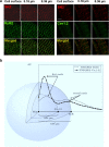
Spatial distribution of SK2, RyR2, and Cav1.2 [108]. a Spatial coupling of SK2, RyR2, and Cav1.2 channels. Stimulated emission depletion (STED) microscopy of SK2, RyR2, and Cav1.2 expression in rabbit ventricular myocytes at three Z planes (scale bar 2 μm). b Visualization of the spatial distribution of SK2, RyR2, and Cav1.2. (AU, arbitrary units; NND, nearest neighbor distance)
Fig. 3
Fig. 3
Functional roles of SK channels in normal and diseased hearts. Distinct roles of SK channels in atria and ventricles are depicted together with remodeling in AF, HF, and calmodulinopathy. EAD, early afterdepolarization; PV, pulmonary veins; AF, atrial fibrillation; VT/VF, ventricular tachycardia and fibrillation; LQTS, long QT syndromes; CPVT, polymorphic ventricular tachycardia; IVF, familial idiopathic ventricular fibrillation
Fig. 4
Fig. 4
Structural modeling of the interactions between CaM mutants and SK2 CaMBD [38]. a Schematic of the 4 α-helices within the C-lobe of CaM. Location of mutations are shown by labels and colored markers. b–d Comparisons of CaMWT (green) and CaMF90L (red, b), CaMF93L (yellow, c), and CaMF142L (purple, d). Side chains of key amino acid residues are shown in stick representation using color scheme shown in panel a. Conformational changes due to CaM mutation are indicated by black arrows in each panel. e C-lobe of apo-CaMWT (colored in green) bound to the C-terminus of hSK2 channel (colored in light brown). Side chains of key amino acids are shown using space-filling representation. f Panel E rotated 90° to the left around the Y-axis. Molecular modeling was performed in Ca2+ free conditions

References

    1. Adelman JP, Maylie J, Sah P. Small-conductance Ca2+-activated K+ channels: form and function. Annu Rev Physiol. 2012;74:245–269. doi: 10.1146/annurev-physiol-020911-153336. - DOI - PubMed
    1. Adsit GS, Vaidyanathan R, Galler CM, Kyle JW, Makielski JC. Channelopathies from mutations in the cardiac sodium channel protein complex. J Mol Cell Cardiol. 2013;61:34–43. doi: 10.1016/j.yjmcc.2013.03.017. - DOI - PMC - PubMed
    1. Allen D, Fakler B, Maylie J, Adelman JP. Organization and regulation of small conductance Ca2+-activated K+ channel multiprotein complexes. J Neurosci. 2007;27:2369–2376. doi: 10.1523/JNEUROSCI.3565-06.2007. - DOI - PMC - PubMed
    1. Azevedo PS, Polegato BF, Minicucci MF, Paiva SAR, Zornoff LAM. Cardiac Remodeling: concepts, clinical impact, pathophysiological mechanisms and pharmacologic treatment. Arq Bras Cardiol. 2016;106:62–69. doi: 10.5935/abc.20160005. - DOI - PMC - PubMed
    1. Benz A, Kossack M, Auth D, Seyler C, Zitron E, Juergensen L, Katus HA, Hassel D. miR-19b Regulates ventricular action potential duration in zebrafish. Sci. Rep. 2016;6:36033. doi: 10.1038/srep36033. - DOI - PMC - PubMed

Publication types

MeSH terms

Substances