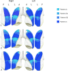
Patients in intensive care unit for COVID-19 pneumonia: the lung ultrasound patterns at admission and discharge. An observational pilot study
- PMID: 33624222
- PMCID: PMC7902088
- DOI: 10.1186/s13089-021-00213-x
Patients in intensive care unit for COVID-19 pneumonia: the lung ultrasound patterns at admission and discharge. An observational pilot study
Erratum in
-
Correction to: Patients in intensive care unit for COVID-19 pneumonia: the lung ultrasound patterns at admission and discharge. An observational pilot study.Ultrasound J. 2021 Mar 9;13(1):15. doi: 10.1186/s13089-021-00219-5. Ultrasound J. 2021. PMID: 33687576 Free PMC article. No abstract available.
Abstract
Background: During COVID-19 pandemic, optimization of the diagnostic resources is essential. Lung Ultrasound (LUS) is a rapid, easy-to-perform, low cost tool which allows bedside investigation of patients with COVID-19 pneumonia. We aimed to investigate the typical ultrasound patterns of COVID-19 pneumonia and their evolution at different stages of the disease.
Methods: We performed LUS in twenty-eight consecutive COVID-19 patients at both admission to and discharge from one of the Padua University Hospital Intensive Care Units (ICU). LUS was performed using a low frequency probe on six different areas per each hemithorax. A specific pattern for each area was assigned, depending on the prevalence of A-lines (A), non-coalescent B-lines (B1), coalescent B-lines (B2), consolidations (C). A LUS score (LUSS) was calculated after assigning to each area a defined pattern.
Results: Out of 28 patients, 18 survived, were stabilized and then referred to other units. The prevalence of C pattern was 58.9% on admission and 61.3% at discharge. Type B2 (19.3%) and B1 (6.5%) patterns were found in 25.8% of the videos recorded on admission and 27.1% (17.3% B2; 9.8% B1) on discharge. The A pattern was prevalent in the anterosuperior regions and was present in 15.2% of videos on admission and 11.6% at discharge. The median LUSS on admission was 27.5 [21-32.25], while on discharge was 31 [17.5-32.75] and 30.5 [27-32.75] in respectively survived and non-survived patients. On admission the median LUSS was equally distributed on the right hemithorax (13; 10.75-16) and the left hemithorax (15; 10.75-17).
Conclusions: LUS collected in COVID-19 patients with acute respiratory failure at ICU admission and discharge appears to be characterized by predominantly lateral and posterior non-translobar C pattern and B2 pattern. The calculated LUSS remained elevated at discharge without significant difference from admission in both groups of survived and non-survived patients.
Conflict of interest statement
The authors declare that they have no competing interests.
Figures


References
-
- Winkler MH, Touw HR, van de Ven PM, Twisk J, Tuinman PR. Diagnostic accuracy of chest radiograph, and when concomitantly studied lung ultrasound, in critically Ill patients with respiratory symptoms: a systematic review and meta-analysis. Crit Care Med. 2018;46:e707–e714. doi: 10.1097/CCM.0000000000003129. - DOI - PubMed
LinkOut - more resources
Full Text Sources
Other Literature Sources
