Review
. 2021 Sep;31(5):1-22.
doi: 10.1002/rmv.2206. Epub 2021 Jan 7.

Innate immune evasion mediated by picornaviral 3C protease: Possible lessons for coronaviral 3C-like protease?

Affiliations
Review

Innate immune evasion mediated by picornaviral 3C protease: Possible lessons for coronaviral 3C-like protease?

Chen Seng Ng et al. Rev Med Virol. 2021 Sep.

Abstract

Severe acute respiratory syndrome coronavirus-2 is the etiological agent of the ongoing pandemic of coronavirus disease-2019, a multi-organ disease that has triggered an unprecedented global health and economic crisis. The virally encoded 3C-like protease (3CLpro ), which is named after picornaviral 3C protease (3Cpro ) due to their similarities in substrate recognition and enzymatic activity, is essential for viral replication and has been considered as the primary drug target. However, information regarding the cellular substrates of 3CLpro and its interaction with the host remains scarce, though recent work has begun to shape our understanding more clearly. Here we summarized and compared the mechanisms by which picornaviruses and coronaviruses have evolved to evade innate immune surveillance, with a focus on the established role of 3Cpro in this process. Through this comparison, we hope to highlight the potential action and mechanisms that are conserved and shared between 3Cpro and 3CLpro . In this review, we also briefly discussed current advances in the development of broad-spectrum antivirals targeting both 3Cpro and 3CLpro .

Keywords: 3CLpro; 3Cpro; Covid-19; SARS-CoV-2; picornaviruses.


Conflict of interest statement

The authors declare that they have no conflict of interest.

Figures

FIGURE 1
FIGURE 1
Crystal structures and superposition of picornaviral 3C protease (3Cpro) and coronaviral 3C‐like protease (3CLpro). (a) Ribbon overlay of the picornaviral 3Cpro structures of poliovirus (PV; PDB 1L1N), foot‐and‐mouth disease virus (FMDV; PDB 2BHG), and human rhinovirus (HRV; PDB 1CQQ). (b) Ribbon overlay of human coronaviral 3CLpro structures of severe acute respiratory syndrome‐coronavirus (SARS‐CoV; PDB 2Q6G), Middle East Respiratory syndrome‐CoV (MERS‐CoV; PDB 4YLU), SARS‐CoV‐2 (PDB 6M2N), and HCoV‐HKU1 (PDB 3D23). (c) A side‐by‐side comparison of PV 3Cpro and SARS‐CoV‐2 3CLpro with the two domains of the chymotrypsin‐like fold highlighted and the active site catalytic residues labeled and highlighted (red). (d) Close‐up images of the active site catalytic residues of PV 3Cpro and SARS‐CoV‐3CLpro are shown
FIGURE 2
FIGURE 2
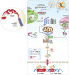
Picornaviruses evade type I interferon immune response via the function of 3C protease (3Cpro). Binding of picornaviruses to their respective receptors facilitates their entry into the cells and release of the 5′‐viral protein genome‐linked‐containing genomic RNA into cytoplasm. Long double‐stranded RNA generated during the replication process binds to MDA5, exposing its CARD and allowing homotypic CARD‐CARD interactions with its downstream adapter, MAVS. Subsequently, MAVS triggers the expression of IFN‐I genes (Ifnb1 and Ifna in dendritic cells) and ISGs for antiviral purposes through the activation of transcription factor IRF3/7 and NF‐κB. To facilitate a robust signaling, more efficient detection of dsRNA occurs in antiviral stress granules. Targets of viral encoded 3Cpro are indicated. CARD, caspase activation and recruitment domain; CTD, C‐terminal binding domain; G3BP1, Ras GTPase‐activating protein‐binding protein 1; IFN‐I, type‐I interferon; IKKε, inhibitor of nuclear factor‐κB (IκB)‐kinase‐ε; IRF3/7, interferon regulatory factors‐3/7; ISGs, interferon‐stimulating genes; MAVS, mitochondrial antiviral signaling protein; MDA5, melanoma‐differentiation associated protein‐5; NF‐κB, nuclear factor‐κB; NEMO, NF‐κB essential modulator; P, phosphate‐group; RIG‐I, retinoic acid‐inducible gene‐I; TBK1, TANK binding kinase‐1; TLR3, Toll‐like receptor 3; TRAF3, TNF‐receptor associated factor‐3; TRIF, Toll/IL‐1 receptor domain‐containing adapter‐inducing interferon‐β
FIGURE 3
FIGURE 3
A proposed mechanistic model for the secondary infection after the initial picornavirus or coronavirus infection. During the first phase of infection, proteins (e.g., TRIP and G3BP1) that are involved in a broad range of host defenses (i.e., against not only RNA viral infection, but also bacterial and DNA viral invasion) are targeted by 3C protease or possibly 3C‐like protease for degradation, rendering the patients more vulnerable to secondary infection. Combinatorial infection during the late phase could result in increased disease severity and mortality
FIGURE 4
FIGURE 4
Picornaviral 3Cpro targets the NLRP3 inflammasome for immune evasion. RNA viruses and other DAMPs could activate NLRP3 inflammasome. Formation of the NLRP3‐dependent inflammasome activates caspase 1, which in turn cleaves pro‐IL‐1β and pro‐IL‐18. GSDMD is also cleaved by caspase 1 and the resulting N‐terminal cleavage products are inserted into the plasma membrane, forming multiple pores and inducing pyroptosis and release of pro‐inflammatory cytokine. Upon picornaviral infection, NLRP3, its upstream signaling proteins (RIP1/RIP3), and its downstream effector GSDMD are all targeted by 3Cpro for degradation. As a result, pyroptosis is inhibited for efficient viral replication. CARD, caspase activation and recruitment domain; DAMPs, danger‐associated molecular patterns; GSDMD, Gasdermin D; LRR, Leucine rich repeat; NLRP3, NLR family and pyrin domain‐containing protein 3; PYD, Pyrin domain; RIP, receptor‐interacting protein; NACHT, NAIP, CIITA, HET‐E and TEP1‐associated families; VPg, viral protein genome‐linked; 3Cpro, 3C protease
FIGURE 5
FIGURE 5
Dispersion of 5′→3′ RNA degradation components within P‐bodies during picornaviral infection. RNA viruses, including picornaviruses, initiate viral replication in a discrete compartment within cytoplasm, generating various RNA species with defined signatures. These includes 5′ppp, 5′p‐ssRNA, dsRNA and viral mRNA. Both picornavirus and coronavirus are able to generate long ssRNA and dsRNA and trigger the translocation of 5’→3′ RNA degradation components, including DCP1, DCP2, and XRN1, into the viral replication complex for degradation of associated viral RNA species. Picornaviral 3Cpro cleaves DCP1, resulting in increased viral particles and infectivity. DCP1/2, decapping protein‐1/2; XRN1, 5’→3′ exoribonuclease‐1; dsRNA, double‐stranded RNA; 3Cpro, 3C protease
FIGURE 6
FIGURE 6
Subversion of host autophagy through picornaviral 3Cpro. Schematic diagram depicted the molecular mechanism for the initiation of host autophagy pathway upon the presence of RNA virus for the clearance of viral‐associated molecules. Picornaviruses utilize its own encoded 2Apro (not shown here) and 3Cpro to cleave key components such as p62, NBR1, SNAP29 and PLEKHM1 to facilitate a more robust replication. ATGs, autophagy‐related genes; DFCP1, double FYVE‐containing protein‐1, FIP200, focal adhesion kinase family interacting protein of 200kD; NBR1, neighbor of BRCA1; SNAP29, synaptosomal‐associated protein‐29; PLEKHM1, Pleckstrin homology and RUN domain containing M1; p62, also known as sequestosome 1 (SQSTM1); STX17, Syntaxin 17; ULK, Unc‐51‐like kinase‐1; UVRAG, UV radiation resistance‐associated gene protein; VAMP8, vesicle‐associated membrane protein‐8; WIPI2, WD‐repeat domain phosphoinositide‐interacting protein‐2; 2Apro, 2A‐protease; 3Cpro, 3C protease
FIGURE 7
FIGURE 7
Schematic workflow of TAILS N‐terminomics screening of 3Cpro and 3CLprosubstrates. Schematic diagram depicts the TAILS workflow and scheme for identification of 3Cpro or 3CLpro substrates. In brief, protein samples from whole cell lysates were subjected to in vitro cleavage by either recombinant purified WT 3Cpro/3CLpro or mutant (C147A) 3Cpro/(C145A) 3CLpro, followed by N‐terminal enrichment using TAILS (left panel). Samples were then combined and subjected to pre‐TAILS shotgun‐like mass‐spectrometry analysis after complete digestion with trypsin. The exposed amine groups of N‐termini generated by the trypsin digestion were then removed by covalently coupling to a high‐molecular weight polyaldehyde polyglycerol polymer. This process allowed for selection via negative enrichment of blocked N termini (middle panel). Peptides were subsequently identified and quantified using high‐resolution mass spectrometry (indicated in the right panel). The resultant high‐confidence candidate substrates were determined through the analysis of the quantified heavy/light (H/L) ratio of dimethylation‐labeled semitryptic neo‐N terminus peptides. They will be subjected to further validation through similar in vitro cleavage assay by 3Cpro/3CLpro, followed by immunoblotting using specific antibodies; TAILS, terminal amine isotopic labeling of substrates; 3Cpro, 3C protease; 3CLpro, 3C‐like protease

Similar articles

Cited by

References

    1. Zhou P, Yang X‐L, Wang X‐G, et al. A pneumonia outbreak associated with a new coronavirus of probable bat origin. Nature. 2020;579:270‐273. - PMC - PubMed
    1. Zhu N, Zhang D, Wang W, et al. A novel coronavirus from patients with pneumonia in China, 2019. N Engl J Med. 2020;382:727‐733. - PMC - PubMed
    1. Coronaviridae Study Group of the International Committee on Taxonomy of Viruses . The species Severe acute respiratory syndrome‐related coronavirus: classifying 2019‐nCoV and naming it SARS‐CoV‐2. Nat Microbiol. 2020;5:536‐544. - PMC - PubMed
    1. Wadman M, Couzin‐Frankel J, Kaiser J, Matacic C. A rampage through the body. Science. 2020;368:356‐360. - PubMed
    1. Huang C, Wang Y, Li X, et al. clinical features of patients infected with 2019 novel coronavirus in Wuhan, China. Lancet. 2020;395:497‐506. - PMC - PubMed

Publication types

Substances