. 2021 Nov;36(13-14):1200-1209.
doi: 10.1177/0883073821993000. Epub 2021 Feb 24.

Enzyme Replacement Therapy for Succinic Semialdehyde Dehydrogenase Deficiency: Relevance in γ-Aminobutyric Acid Plasticity

Affiliations

Enzyme Replacement Therapy for Succinic Semialdehyde Dehydrogenase Deficiency: Relevance in γ-Aminobutyric Acid Plasticity

Henry Hing Cheong Lee et al. J Child Neurol. 2021 Nov.

Abstract

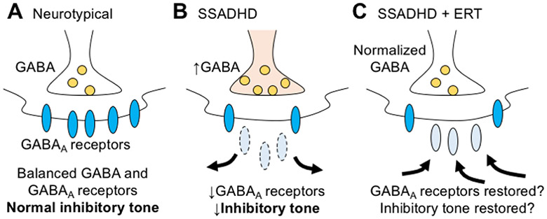
Succinic semialdehyde dehydrogenase deficiency (SSADHD) is a rare inborn metabolic disorder caused by the functional impairment of SSADH (encoded by the ALDH5A1 gene), an enzyme essential for metabolism of the inhibitory neurotransmitter γ-aminobutyric acid (GABA). In SSADHD, pathologic accumulation of GABA and its metabolite γ-hydroxybutyrate (GHB) results in broad spectrum encephalopathy including developmental delay, ataxia, seizures, and a heightened risk of sudden unexpected death in epilepsy (SUDEP). Proof-of-concept systemic SSADH restoration via enzyme replacement therapy increased survival of SSADH knockout mice, suggesting that SSADH restoration might be a viable intervention for SSADHD. However, before testing enzyme replacement therapy or gene therapy in patients, we must consider its safety and feasibility in the context of early brain development and unique SSADHD pathophysiology. Specifically, a profound use-dependent downregulation of GABAA receptors in SSADHD indicates a risk that any sudden SSADH restoration might diminish GABAergic tone and provoke seizures. In addition, the tight developmental regulation of GABA circuit plasticity might limit the age window when SSADH restoration is accomplished safely. Moreover, given SSADH expressions are cell type-specific, targeted instead of global restoration might be necessary. We therefore describe 3 key parameters for the clinical readiness of SSADH restoration: (1) rate, (2) timing, and (3) cell type specificity. Our work focuses on the construction of a novel SSADHD mouse model that allows "on-demand" SSADH restoration for the systematic investigation of these key parameters. We aim to understand the impacts of specific SSADH restoration protocols on brain physiology, accelerating bench-to-bedside development of enzyme replacement therapy or gene therapy for SSADHD patients.

Keywords: epilepsy; genetics; inborn errors of metabolism; metabolism; mitochondrial disorder; neurodevelopment; seizures; status epilepticus; treatment.


Figures

Fig. 1.
Fig. 1.
γ-aminobutyric acid (GABA) metabolic pathway. Cytosolic glutamate is converted by glutamic acid decarboxylase (GAD) to form GABA, which is subsequently translocated into the mitochondria, where GABA is reversibly converted by GABA transaminase (GABA-T) to succinic semialdehyde (SSA). SSA is converted either by SSA reductase (SSAR) to γ-hydroxybutyric acid (GHB), or by SSA dehydrogenase (SSADH) to succinate, which then enters the Krebs cycle. In the absence of SSADH (i.e., SSADH deficiency), GABA and GHB are accumulated to pathologic levels.
Fig. 2.
Fig. 2.
Use-dependent compensatory GABAA receptor expression underlies seizures in SSADHD and potential enzyme replacement therapy (ERT) response. (A) Under neurotypical situation, balanced levels of GABA and GABAA receptors result in normal inhibitory tone. (B) In SSADHD, pathologic accumulation of GABA leads to use-dependent reduction of GABAA receptors. Despite a hyper-GABAergic condition, overall inhibitory tone is sufficiently impaired, resulting in seizures in SSADHD patients. (C) ERT in SSADHD normalizes (reduces) GABA levels in a setting of reduced GABAA receptors. Successful ERT outcomes depends on plastic restoration of functional GABAA receptors and inhibitory tone.
Fig. 3.
Fig. 3.
Construction of the aldh5a1lox-rtTA-STOP mouse. (A) The endogenous aldh5a1 gene is disrupted by CRISPR/Cas9-mediated homology directed repair in its first intron with the insertion of a gene cassette containing a splice acceptor (AG) and the rtTA-STOP sequence flanked by two loxP sites. (B) At baseline, aldh5a1lox-rtTA-STOP mice are SSADH-null due to the disrupted aldh5a1 gene. Instead, rtTA expression is driven by endogenous aldh5a1 promoter activities (to combine with a second mouse, TRE-aldh5a1, for doxycycline-mediated rescue strategy; see Fig. 7 for details). (C) Upon Cre-recombination, aldh5a1 is reconstituted for re-expression (aldh5a1Δ).
Fig. 4.
Fig. 4.
Experimental paradigms studying the impacts of rate of SSADH restoration in mice. Same total amount of AAV will be injected across different timespans (1, 3 or 5 days) to represent gradual (A), moderate (B) and abrupt (C) SSADH restoration.
Fig. 5.
Fig. 5.
Experimental paradigms studying the impacts of age of SSADH restoration in mice. Same total amount of AAV will be injected at different ages (P5 or P15) to represent pre-symptomatic (A) and peri-symptomatic (B) SSADH restoration.
Fig. 6.
Fig. 6.
Transcripts expression of SSADH. (A) In situ hybridization (ISH) data of aldh5a1 transcripts in adult (P56) C57Bl/6J mouse brain. Credit: Allen Brain Institute online database (http://mouse.brain-map.org/). Note the brain-wide expression of aldh5a1, and its enhanced expression in the hippocampus and the cerebellum. (B) Single-cell RNAseq data of aldh5a1 in mouse cortex and hippocampus. Credit: The Linnarsson lab (http://linnarssonlab.org/cortex/).. Cell types are classified as interneurons and pyramidal cells (PYR). (C) Single-cell RNAseq data of aldh5a1 in the mouse whole cortex and the hippocampus. Credit: Allen Brain Institute online database (https://celltypes.brain-map.org/rnaseq/mouse_ctx-hip_10x). Single-cell RNAseq data in (B) and (C) suggested that aldh5a1 expression is biased towards GABAergic interneurons. Molecular markers of interneuron subclass are included for comparison. VIP, vasoactive intestinal polypeptide; SOM, somatostatin; PV, parvalbumin.
Fig. 7.
Fig. 7.
Conceptual design of a reversible SSADH mouse model. Breeding aldh5a1lox-rtTA-STOP and TRE-aldh5a1 mice allows reversible expression of recombinant aldh5a1 in the presence of doxycycline (Dox) tightly driven by a Tet-responsive element (TRE).
Fig. 8.
Fig. 8.
Rate-dependent GFP expression via AAV-PHP.B injected across various timespans. Representative confocal micrographs showing the cerebellum (A1-2, B1-2 and C1-2) and the hippocampus (A3-4, B3-4 and C3-4) at 7 and 14 days post-injection (d.p.i.), in low magnification (10X), using escalating doses of AAV-PHP.B to mimic gradual (A), moderate (B) and rapid (C) transgene expression across various timespans (E). High magnification (40X) of individual neurons in selected brain regions in B are shown in D. Quantification is presented in F. Scale bars: 500μm (A1, A3), 20μm (D1). Note rate-dependent expression at 7 d.p.i across these three dosing schedules (L to R: gradual, moderate and rapid), but these changes are largely diminished at 14 d.p.i. N=4 (per dosing schedule and post-injection time point) from two independent experiments. Statistical analysis: One-way ANOVA followed by Bonferroni’s Multiple Comparison Test. *P<0.05, ***P<0.001, n.s. = not significant.
Fig. 9.
Fig. 9.
AAV-PHP.B intraperitoneally (IP) injected at P10 transduced various interneuron cell types in the brain. Representative confocal micrographs of cryopreserved brain sections showing AAV-PHP.B-CAG-GFP transduced cells (top row in green) in the hippocampus and the cerebellum. Immunostaining was performed using various interneuron cellular markers (middle row in red). Arrow heads indicate GFP-expressing cells co-immunostained by respective interneuron cellular markers (bottom row). Selected identified GFP+ cells are shown in high magnification in insets. Scale bar: 50μm. VIP, vasoactive intestinal polypeptide-expressing interneurons; PV, parvalbumin.

References

    1. Gibson KM et al. Succinic semialdehyde dehydrogenase deficiency: an inborn error of gamma-aminobutyric acid metabolism. Clin Chim Acta 133, 33–42, doi:10.1016/0009-8981(83)90018-9 (1983). - DOI - PubMed
    1. Pearl PL et al. Inherited disorders of gamma-aminobutyric acid metabolism and advances in ALDH5A1 mutation identification. Dev Med Child Neurol 57, 611–617, doi:10.1111/dmcn.12668 (2015). - DOI - PMC - PubMed
    1. Jakobs C et al. Urinary excretion of gamma-hydroxybutyric acid in a patient with neurological abnormalities. The probability of a new inborn error of metabolism. Clin Chim Acta 111, 169–178, doi:10.1016/0009-8981(81)90184-4 (1981). - DOI - PubMed
    1. Pearl PL, Wiwattanadittakul N, Roullet JB & Gibson KM in GeneReviews((R)) (eds Adam MP et al.) (1993).
    1. Malaspina P et al. Succinic semialdehyde dehydrogenase deficiency (SSADHD): Pathophysiological complexity and multifactorial trait associations in a rare monogenic disorder of GABA metabolism. Neurochem Int 99, 72–84, doi:10.1016/j.neuint.2016.06.009 (2016). - DOI - PMC - PubMed

Publication types

MeSH terms

Substances

Supplementary concepts