

Soluble fms-like tyrosine kinase-1 and angiotensin2 target calcitonin gene-related peptide family peptides in maternal vascular smooth muscle cells in pregnancy†
- PMID: 33624744
- PMCID: PMC8111240
- DOI: 10.1093/biolre/ioab026
Soluble fms-like tyrosine kinase-1 and angiotensin2 target calcitonin gene-related peptide family peptides in maternal vascular smooth muscle cells in pregnancy†
Abstract
Calcitonin gene-related peptide (CALCB), adrenomedullin (ADM), and adrenomedullin2 (ADM2) are hypotensive peptides that belong to CALCB family of peptides. Goal of this study was to identify the effect of fms-like tyrosine kinase (sFLT-1) and angiotensin2 (Ang2) on the function of these peptides in OA smooth muscle cells (OASMC) and assess the sensitivity of OA for these peptides in preeclampsia (PE) and normotensive pregnancy.
Methods: Peptide function was assessed by Cyclic adenosine monophosphate (cAMP) assays and wire myograph; mRNA expression by Polymerase chain reaction (PCR) and protein-protein interaction by proximity ligation assay and co-immunoprecipitation.
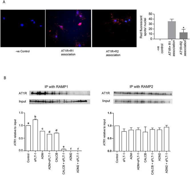
Findings: All three peptides increased cAMP synthesis in the order of efficacy CALCB > ADM = ADM2 and vascular endothelial growth factor (VEGF) mRNA in OASMC (P < 0.05); sFLT-1 mediated decrease in cAMP synthesis (P < 0.05) is differentially rescued by all three CALCB family peptides in OASMC (P < 0.005); sFLT-1 decreased receptor activity-modifying protein (RAMP)1 and RAMP2 mRNA expression (P < 0.05); Ang2 decreased the expression of calcitonin-receptor-like receptor and RAMP1 mRNA and desensitized CALCB and ADM2 receptors in OASMC (P < 0.05); sFLT-1 increased RAMP1and Ang2 type 1 receptor (AT1R) interaction in OASMC which is inhibited in presence of all three peptides; and all three peptides relax OA in PE with enhanced ADM2 response (P < 0.05).
Conclusion: sFLT-1 and Ang2 impair OASMC mediated functional responses of CALCB family peptides which can be inhibited by respective peptide treatment. The sensitivity of OA for CALCB, ADM, and ADM2-mediated relaxation is retained in PE.
Keywords: angiotensin 2; cAMP; calcitonin gene-related peptides; pregnancy; sFLT-1; vascular smooth muscle cells.
© The Author(s) 2021. Published by Oxford University Press on behalf of Society for the Study of Reproduction. All rights reserved. For permissions, please e-mail: journals.permissions@oup.com.
Figures


Similar articles
-
Calcitonin Gene Related Peptide, Adrenomedullin, and Adrenomedullin 2 Function in Uterine Artery During Human Pregnancy.Endocrinology. 2022 Jan 1;163(1):bqab204. doi: 10.1210/endocr/bqab204. Endocrinology. 2022. PMID: 34558598 Free PMC article.
-
Calcitonin Gene-Related Peptide Rescues Proximity Associations of Its Receptor Components, Calcitonin Receptor-Like Receptor and Receptor Activity-Modifying Protein 1, in Rat Uterine Artery Smooth Muscle Cells Exposed to Tumor Necrosis Factor Alpha.Biol Reprod. 2016 Dec;95(6):126. doi: 10.1095/biolreprod.116.143529. Epub 2016 Oct 26. Biol Reprod. 2016. PMID: 27784654 Free PMC article.
-
Involvement of Receptor Activity-Modifying Protein 3 (RAMP3) in the Vascular Actions of Adrenomedullin in Rat Mesenteric Artery Smooth Muscle Cells.Biol Reprod. 2015 Nov;93(5):116. doi: 10.1095/biolreprod.115.134585. Epub 2015 Sep 30. Biol Reprod. 2015. PMID: 26423127 Free PMC article.
-
Nutraceutical Targeting of Placental Synthesis of Soluble Fms-Like Tyrosine Kinase- 1 (sFlt-1) as Strategy for Preventing and Controlling Pre-eclampsia.Curr Pharm Des. 2018;24(20):2255-2263. doi: 10.2174/1381612824666180723162327. Curr Pharm Des. 2018. PMID: 30039754 Review.
-
Calcitonin gene-related family peptides in vascular adaptations, uteroplacental circulation, and fetal growth.Curr Vasc Pharmacol. 2013 Sep;11(5):641-54. doi: 10.2174/1570161111311050007. Curr Vasc Pharmacol. 2013. PMID: 24063381 Review.
Cited by
-
Integrated pan-cancer analysis of ADM's role in prognosis, immune modulation and resistance.Front Immunol. 2025 Jun 3;16:1573250. doi: 10.3389/fimmu.2025.1573250. eCollection 2025. Front Immunol. 2025. PMID: 40529377 Free PMC article.
-
Calcitonin Gene Related Peptide, Adrenomedullin, and Adrenomedullin 2 Function in Uterine Artery During Human Pregnancy.Endocrinology. 2022 Jan 1;163(1):bqab204. doi: 10.1210/endocr/bqab204. Endocrinology. 2022. PMID: 34558598 Free PMC article.
-
Role of adrenomedullin2/ intermedin in pregnancy induced vascular and metabolic adaptation in mice.Front Physiol. 2023 Feb 17;14:1116042. doi: 10.3389/fphys.2023.1116042. eCollection 2023. Front Physiol. 2023. PMID: 36875025 Free PMC article.
-
Calcitonin gene-related peptide protects from soluble fms-like tyrosine kinase-1-induced vascular dysfunction in a preeclampsia mouse model.Front Physiol. 2023 Aug 31;14:1221684. doi: 10.3389/fphys.2023.1221684. eCollection 2023. Front Physiol. 2023. PMID: 37719463 Free PMC article.
References
-
- Chauhan M, Yallampalli U, Reed L, Yallampalli C. Adrenomedullin 2 antagonist infusion to rats during midgestation causes fetoplacental growth restriction through apoptosis. Biol Reprod 2006; 75:940–947. - PubMed
-
- McLatchie LM, Fraser NJ, Main MJ, Wise A, Brown J, Thompson N, Solari R, Lee MG, Foord SM. RAMPs regulate the transport and ligand specificity of the calcitonin-receptor-like receptor. Nature 1998; 393:333–339. - PubMed
-
- Yallampalli C, Chauhan M, Sathishkumar K. Calcitonin gene-related family peptides in vascular adaptations, uteroplacental circulation, and fetal growth. Curr Vasc Pharmacol 2013; 11:641–654. - PubMed
-
- Penchalaneni J, Wimalawansa SJ, Yallampalli C. Adrenomedullin antagonist treatment during early gestation in rats causes fetoplacental growth restriction through apoptosis. Biol Reprod 2004; 71:1475–1483. - PubMed
-
- Yallampalli C, Chauhan M, Endsley J, Sathishkumar K. Calcitonin gene related family peptides: importance in normal placental and fetal development. Adv Exp Med Biol 2014; 814:229–240. - PubMed
Publication types
MeSH terms
Substances
Grants and funding
LinkOut - more resources
Full Text Sources
Other Literature Sources
Molecular Biology Databases
Miscellaneous
