Case Reports
. 2021 Oct 1;41(10):2106-2114.
doi: 10.1097/IAE.0000000000003153.

BACILLARY LAYER DETACHMENT BECAUSE OF MACULAR NEOVASCULARIZATION

Affiliations
Case Reports

BACILLARY LAYER DETACHMENT BECAUSE OF MACULAR NEOVASCULARIZATION

Jesse J Jung et al. Retina. .

Abstract

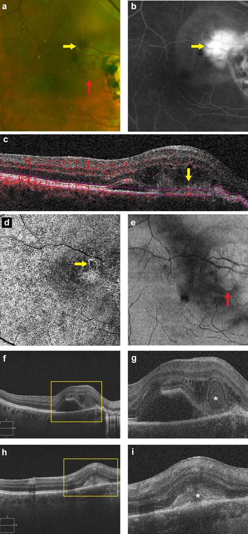
Purpose: To describe the clinical and multimodal imaging features of bacillary layer detachment (BD), and its response to intravitreal anti-vascular endothelial growth factor therapy, in eyes with macular neovascularization.

Methods: Retrospective, observational case series of 14 eyes (14 patients, 7 men) imaged with eyes (14 patients, 7 men) were imaged with spectral-domain optical coherence tomography, and either fluorescein angiography or optical coherence tomography angiography. Therapeutic response was monitored with serial imaging and best-corrected visual acuity assessments.

Results: The mean age was 75 ± 13 (range: 45-96) years, with mean follow-up duration of 27 ± 21 (range: 1-56) months. Neovascular age-related macular degeneration was found in 71% (10/14) eyes. Type 2 macular neovascularization lesions were associated with BD in all 14 eyes. Subretinal hemorrhage was noted in 79% (11/14) eyes. BD promptly resolved after intravitreal antivascular endothelial growth factor therapy in all eyes. The baseline best-corrected visual acuity improved from logarithm of the minimum angle of resolution 0.84 ± 0.32 (Snellen equivalent 20/138) to logarithm of the minimum angle of resolution 0.48 ± 0.31 (Snellen equivalent 20/60) at the last follow-up, with treatment of the macular neovascularization.

Conclusion: Type 2 macular neovascularization and subretinal hemorrhage are associated with BDs, which may be due to a rapid influx of exudative fluid into the potential space between the external limiting membrane and ellipsoid zone. Intravitreal antivascular endothelial growth factor therapy results in rapid resolution of BDs and visual improvement in most eyes.


Figures

Figure 1.
Figure 1.. Representative Spectral-Domain and Swept-Source Optical Coherence Tomography Examples of Bacillary Layer Detachment in all 14 Cases.
Spectral-domain or swept-source-optical coherence tomography near-infrared and corresponding structural B-scans demonstrating bacillary layer detachment (BD) in each of the 14 eyes included in this study. Image numbering corresponds to subject number presented in Table 1. Yellow box with magnifed insert of area of interest with yellow arrows indicating the location of BD in each eye.
Figure 2.
Figure 2.. Multimodal Imaging of Bacillary Layer Detachment in a case of Macular Telangiectasia Type 2.
For case #9, the macula was imaged at baseline with the following modalities to highlight the bacillary layer detachment (BD) and adjacent Type 2 macular neovascularization (MNV) with subretinal hyperreflective exudative material: a) Color fundus photography; b) Green-wavelength fundus autofluorescence; c and d) Pair of fluorescein angiography images obtained at early and late timepoints respectively, demonstrating early leakage suggestive of type 2 MNV; e) Spectral-domain optical coherence tomography angiography 3×3 mm en-face image demonstrating a subretinal MNV with f) corresponding segmentation on structural B-scan; g) Magnified view of spectral-domain optical coherence tomography B-scan demonstrating a BD adjacent to a type 2 CNV, with white arrowheads indicating elevated external limiting membrane anteriorly and white asterisk indicating the intracystic space with hyperreflective exudation in the posterior portion of the BD.
Figure 3.
Figure 3.. Multimodal Imaging and Treatment Response of Bacillary Layer Detachment in a case with Neovascular Age-Related Macular Degeneration.
Case #2 was diagnosed with bacillary layer detachment (BD) secondary to type 2 macular neovascularization (MNV), attributable to inflammatory ampiginous chorioretinitis. a) Color fundus photography demonstrates an area of pigment deposition and subretinal hemorrhage corresponding to the type 2 MNV (yellow arrow), and an intraretinal cyst corresponding to the BD (red arrow); b) Fluorescein angiography demonstrates early hyperfluorescence and leakage suggestive of type 2 MNV within the extrafoveal, peripapillary zone (yellow arrow), which can also be visualized on panel ‘c’. Spectral-domain optical coherence tomography angiography (SD-OCTA) structural B-scan with segmentation lines, as well as the corresponding d) SD-OCTA 6×6 mm en-face scan acquired at the subretinal layer (yellow arrow), and e) SD-OCT en-face c-scan showing a circular area with hyperreflective border (red arrow) corresponding to the BD. Panels f and g correspond to SD-OCT structural B-scan demonstrating BD (magnified view of the yellow box, ‘g’, with BD indicated by white asterisk). Six weeks after treatment with a single injection of bevacizumab 1.25mg/0.05 ml (Genentech Inc., South San Francisco, CA, USA) (panel h, magnified view of the yellow box in ‘i’), the Type 2 MNV has consolidated into a more Type 1 appearing MNV (white asterisk) with an enveloping retinal pigment epithelal layer and the BD has entirely resolved, with visual acuity improving from 20/80 to 20/25.
Figure 4.
Figure 4.. Multimodal Imaging and Treatment Response of Bacillary Layer Detachment in a case of inflammatory posterior uveitis.
Case #3 was diagnosed with neovascular age-related macular degeneration (nAMD) and a bacillary layer detachment (BD) due to type 2 macular neovascularization (MNV). a) Color fundus photography demonstrates the presence of subretinal hemorrhage and an intraretinal cyst at the juxtafoveal region (yellow arrow); b) Fluorescein angiography demonstrates early hyperfluorescence and leakage suggestive of type 2 MNV (yellow arrow); c) Near-infrared reflectance image demonstrates a circular intraretinal cyst corresponding to the BD centrally; d) Swept-source optical coherence tomography B-scan demonstrating BD at baseline (magnified view of the yellow box in panel ‘e’, with BD indicated by white asterisk); f) 4 weeks after treatment with a single injection of bevacizumab 1.25mg/0.05 ml (Genentech Inc., South San Francisco, CA, USA), the BD resolved entirely, with VA improving from 20/160 to 20/50.

Comment in

  • Reply.
    Soh YQ, Hoang QV, Freund KB, Jung JJ. Soh YQ, et al. Retina. 2022 Jan 1;42(1):e1-e3. doi: 10.1097/IAE.0000000000003240. Retina. 2022. PMID: 34173360 Free PMC article.
  • Correspondence.
    Mambretti M, Casalino G. Mambretti M, et al. Retina. 2022 Jan 1;42(1):e1. doi: 10.1097/IAE.0000000000003239. Retina. 2022. PMID: 34173363 No abstract available.

References

    1. Spaide RF, Curcio CA. Anatomical correlates to the bands seen in the outer retina by optical coherence tomography: literature review and model. Retina. 2011;31(8):1609–1619. - PMC - PubMed
    1. Ross DH, Clark ME, Godara P, et al.RefMoB, a Reflectivity Feature Model-Based Automated Method for Measuring Four Outer Retinal Hyperreflective Bands in Optical Coherence Tomography. Invest Ophthalmol Vis Sci. 2015;56(8):4166–4176. - PMC - PubMed
    1. Jonnal RS, Kocaoglu OP, Zawadzki RJ, Lee S-H, Werner JS, Miller DT. The cellular origins of the outer retinal bands in optical coherence tomography images. Invest Ophthalmol Vis Sci. 2014;55(12):7904–7918. - PMC - PubMed
    1. Drexler W, Morgner U, Ghanta RK, Kärtner FX, Schuman JS, Fujimoto JG. Ultrahigh-resolution ophthalmic optical coherence tomography. Nat Med. 2001;7(4):502–507. - PMC - PubMed
    1. Cuenca N, Ortuño-Lizarán I, Pinilla I. Cellular Characterization of OCT and Outer Retinal Bands Using Specific Immunohistochemistry Markers and Clinical Implications. Ophthalmology. 2018;125(3):407–422. - PubMed

Publication types

MeSH terms

Substances

LinkOut - more resources