Case Reports
. 2021 Jul;300(1):E296-E300.
doi: 10.1148/radiol.2021210275. Epub 2021 Feb 24.

Lymphadenopathy in COVID-19 Vaccine Recipients: Diagnostic Dilemma in Oncologic Patients

Affiliations
Case Reports

Lymphadenopathy in COVID-19 Vaccine Recipients: Diagnostic Dilemma in Oncologic Patients

Can Özütemiz et al. Radiology. 2021 Jul.

Abstract

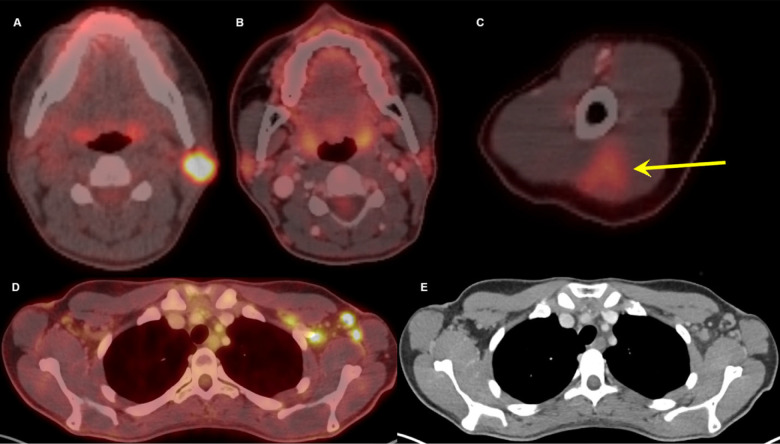
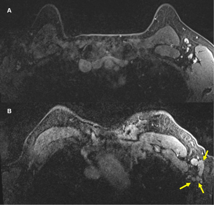
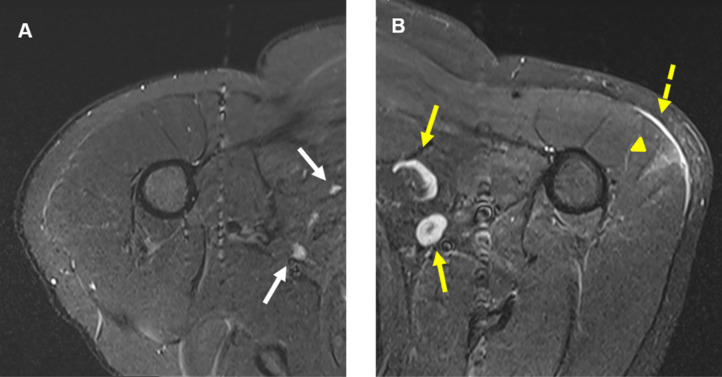
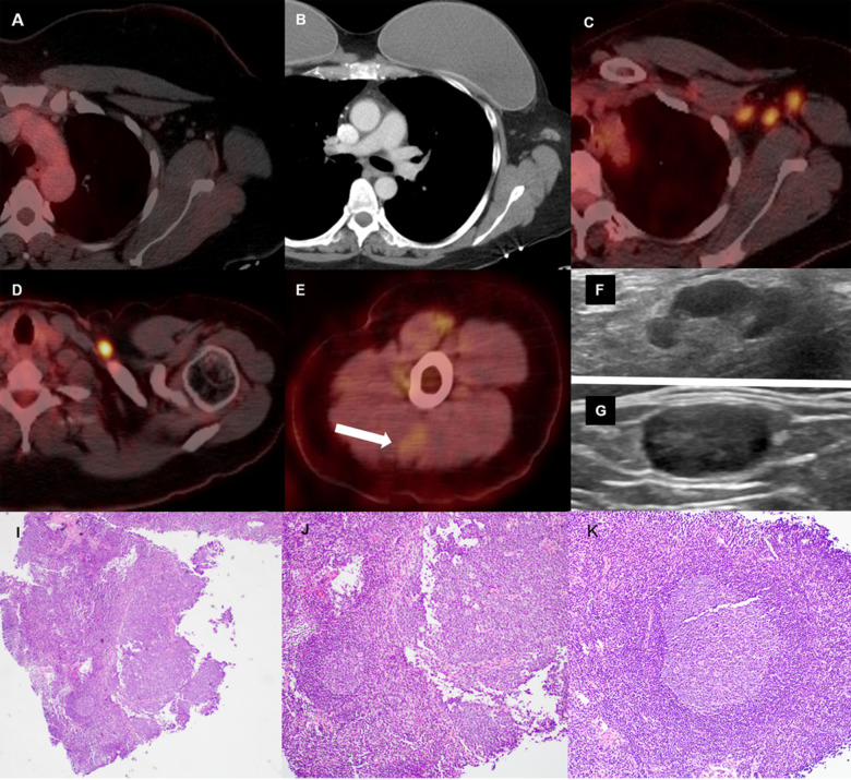
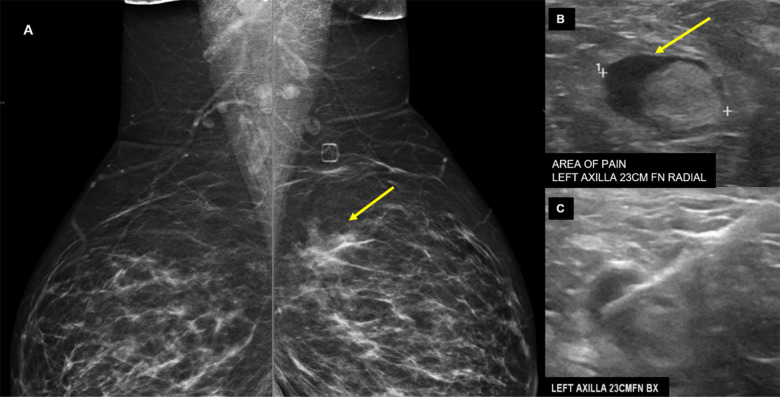
Five cases of axillary lymphadenopathy are presented, which occurred after COVID-19 vaccination and mimicked metastasis in a vulnerable oncologic patient group. Initial radiologic diagnosis raised concerns for metastasis. However, further investigation revealed that patients received COVID-19 vaccinations in the ipsilateral arm prior to imaging. In two cases, lymph node biopsy results confirmed vaccination-related reactive lymphadenopathy. Ipsilateral axillary swelling or lymphadenopathy was reported based on symptoms and physical examination in COVID-19 vaccine trials. Knowledge of the potential for COVID-19 vaccine-related ipsilateral adenopathy is necessary to avoid unnecessary biopsy and change in therapy. © RSNA, 2021.


Figures

Thirty-two-year-old female. A, Axial fused 18-FDG PET/CT showed hypermetabolic biopsy proven intraparotid lymph node with metastatic malignant melanoma. B, Three-month follow-up axial fused 18-FDG PET/CT shows complete resolution of the neck mass following chemotherapy, C, while left arm shows hypermetabolic triangular shaped inflammation (arrow) at the COVID vaccine injection site. D, Axial fused images at the axilla level shows multiple new hypermetabolic lymph nodes. E, Axial contrast enhanced CT demonstrates mild fat stranding surrounding the ovoid lymph nodes with preserved fatty hilum.
Figure 1:
Thirty-two-year-old female. A, Axial fused 18-FDG PET/CT showed hypermetabolic biopsy proven intraparotid lymph node with metastatic malignant melanoma. B, Three-month follow-up axial fused 18-FDG PET/CT shows complete resolution of the neck mass following chemotherapy, C, while left arm shows hypermetabolic triangular shaped inflammation (arrow) at the COVID vaccine injection site. D, Axial fused images at the axilla level shows multiple new hypermetabolic lymph nodes. E, Axial contrast enhanced CT demonstrates mild fat stranding surrounding the ovoid lymph nodes with preserved fatty hilum.
57-year-old female. A, Axial contrast enhanced fat saturated T1 sequence of MRI from a year ago showed sub-centimeter, benign lymph nodes in the left axilla. B, Most recent contrast enhanced axial fat saturated T1 sequence at approximately same level showed multiple new, enlarged lymph nodes (yellow arrows) in the left axilla. There were no other suspicious findings to suggest a breast cancer primary.
Figure 2:
57-year-old female. A, Axial contrast enhanced fat saturated T1 sequence of MRI from a year ago showed sub-centimeter, benign lymph nodes in the left axilla. B, Most recent contrast enhanced axial fat saturated T1 sequence at approximately same level showed multiple new, enlarged lymph nodes (yellow arrows) in the left axilla. There were no other suspicious findings to suggest a breast cancer primary.
41-year-old male. A, Axial T2-STIR MRI of the right shoulder shows normal looking muscular and osseous structures, and normal lymph nodes (white arrows). B, Axial T2-STIR MRI of the left shoulder correspondent to the level of A, shows new subcutaneous edema (dashed yellow arrow) overlying a wedge-shaped intramuscular edema (yellow arrowhead) in the left deltoid muscle. In addition, there are multiple prominent ovoid shaped lymph nodes (yellow arrows) in the ipsilateral axilla, asymmetric to the right and new compared with prior MRIs (not shown).
Figure 3:
41-year-old male. A, Axial T2-STIR MRI of the right shoulder shows normal looking muscular and osseous structures, and normal lymph nodes (white arrows). B, Axial T2-STIR MRI of the left shoulder correspondent to the level of A, shows new subcutaneous edema (dashed yellow arrow) overlying a wedge-shaped intramuscular edema (yellow arrowhead) in the left deltoid muscle. In addition, there are multiple prominent ovoid shaped lymph nodes (yellow arrows) in the ipsilateral axilla, asymmetric to the right and new compared with prior MRIs (not shown).
46-year-old female with triple negative left breast cancer, disease free for three years. A, Axial fused 18-FDG PET/CT three years earlier with no concerning lymph node in the left axilla. B, Surveillance contrast enhanced axial chest CT showed new left axillary lymphadenopathy with fat stranding 15 days after the first Covid-19 vaccine. Further evaluation with PET/CT six days after the 2nd dose of vaccine, demonstrated, C, multiple enlarged hypermetabolic left axillary lymph nodes and, D, a hypermetabolic round shaped left supraclavicular lymph node in axial fused 18-FDG PET/CT images. E, A subtle wedge-shaped intramuscular hypermetabolism (white arrow) was also noted in this case, similar to first and third cases. Ultrasonography guided core needle biopsy was performed. F, On ultrasonography, axillary lymph nodes had thickened cortex while the supraclavicular lymph node demonstrated, G, thickened cortex with loss of normal fatty hilum. I, Hematoxylin and eosin staining under 40x magnification shows enlarged germinal center with interfollicular expansion by small lymphocytes. 100x magnification images show, J, prominent germinal center with tingible body macrophage and, K, reactive germinal center with expansion of interfollicular regions by small lymphocytes and focally prominent endothelial cells.
Figure 4:
46-year-old female with triple negative left breast cancer, disease free for three years. A, Axial fused 18-FDG PET/CT three years earlier with no concerning lymph node in the left axilla. B, Surveillance contrast enhanced axial chest CT showed new left axillary lymphadenopathy with fat stranding 15 days after the first Covid-19 vaccine. Further evaluation with PET/CT six days after the 2nd dose of vaccine, demonstrated, C, multiple enlarged hypermetabolic left axillary lymph nodes and, D, a hypermetabolic round shaped left supraclavicular lymph node in axial fused 18-FDG PET/CT images. E, A subtle wedge-shaped intramuscular hypermetabolism (white arrow) was also noted in this case, similar to first and third cases. Ultrasonography guided core needle biopsy was performed. F, On ultrasonography, axillary lymph nodes had thickened cortex while the supraclavicular lymph node demonstrated, G, thickened cortex with loss of normal fatty hilum. I, Hematoxylin and eosin staining under 40x magnification shows enlarged germinal center with interfollicular expansion by small lymphocytes. 100x magnification images show, J, prominent germinal center with tingible body macrophage and, K, reactive germinal center with expansion of interfollicular regions by small lymphocytes and focally prominent endothelial cells.
38-year-old presented for evaluation of left breast and left axillary pain. A, Bilateral MLO views demonstrate an asymmetry (yellow arrow) in the left superior breast which was biopsied as pseudoangiomatous stromal hyperplasia. B, Ultrasound evaluation of the left axilla demonstrates a lymph node with abnormally thickened cortex (yellow arrow). C, Ultrasound guided core needle biopsy for lymph node revealed reactive follicular hyperplasia.
Figure 5:
38-year-old presented for evaluation of left breast and left axillary pain. A, Bilateral MLO views demonstrate an asymmetry (yellow arrow) in the left superior breast which was biopsied as pseudoangiomatous stromal hyperplasia. B, Ultrasound evaluation of the left axilla demonstrates a lymph node with abnormally thickened cortex (yellow arrow). C, Ultrasound guided core needle biopsy for lymph node revealed reactive follicular hyperplasia.

References

    1. Moderna COVID-19 Vaccine . US Food & Drug Administration Web site. https://www.fda.gov/emergency-preparedness-and-response/coronavirus-dise.... Accessed February 06, 2021.
    1. Pfizer-BioNTech COVID-19 Vaccine . US Food & Drug Administration Web site. https://www.fda.gov/emergency-preparedness-and-response/coronavirus-dise.... Accessed February 06, 2021.
    1. Covid data tracker . US Center for Disease Control and Prevention Web site. https://covid.cdc.gov/covid-data-tracker/#vaccinations. Accessed February 06, 2021.
    1. Local Reactions, Systemic Reactions, Adverse Events, and Serious Adverse Events: Moderna COVID-19 Vaccine . US Center for Disease Control and Prevention Web site. https://www.cdc.gov/vaccines/covid-19/info-by-product/moderna/reactogeni.... Accessed February 06, 2021.
    1. Polack FP, Thomas SJ, Kitchin N, Absalon J, Gurtman A, Lockhart S, Perez JL, Perez Marc G, Moreira ED, Zerbini C, Bailey R, Swanson KA, Roychoudhury S, Koury K, Li P, Kalina WV, Cooper D, Frenck RW, Jr., Hammitt LL, Tureci O, Nell H, Schaefer A, Unal S, Tresnan DB, Mather S, Dormitzer PR, Sahin U, Jansen KU, Gruber WC, Group CCT. Safety and Efficacy of the BNT162b2 mRNA Covid-19 Vaccine. N Engl J Med 2020;383:2603-2615. doi: 10.1056/NEJMoa2034577 - PMC - PubMed

Publication types

Substances

LinkOut - more resources