. 2021 Jul 2;33(5):1706-1727.
doi: 10.1093/plcell/koab061.

The state of oligomerization of Rubisco controls the rate of synthesis of the Rubisco large subunit in Chlamydomonas reinhardtii

Affiliations

The state of oligomerization of Rubisco controls the rate of synthesis of the Rubisco large subunit in Chlamydomonas reinhardtii

Wojciech Wietrzynski et al. Plant Cell. .

Abstract

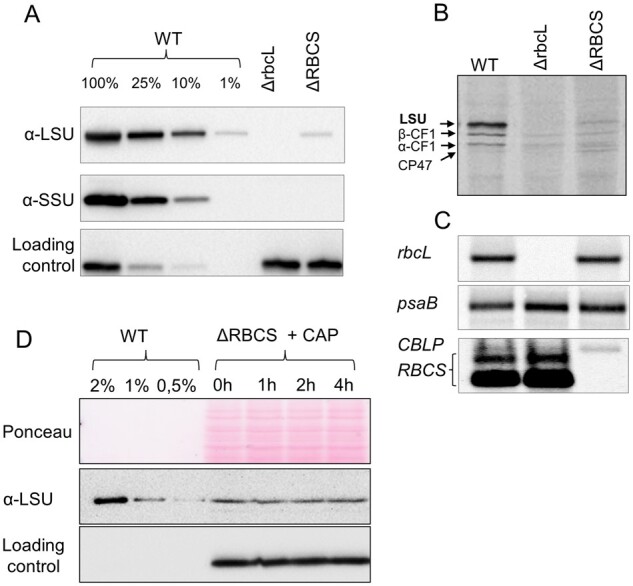
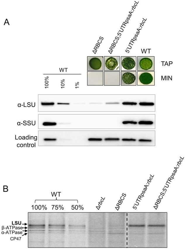
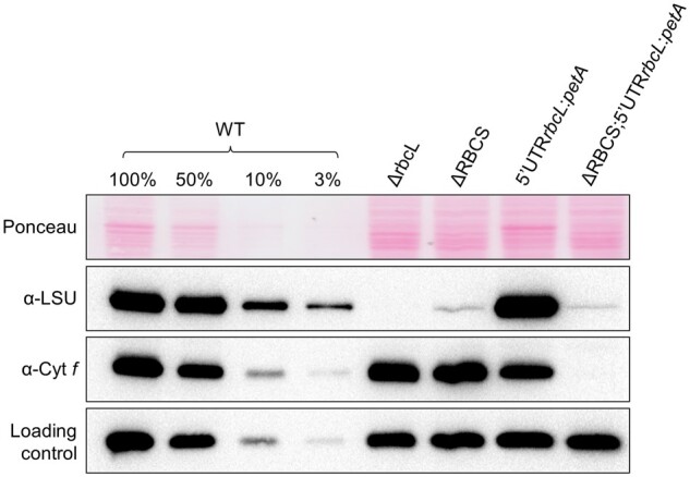
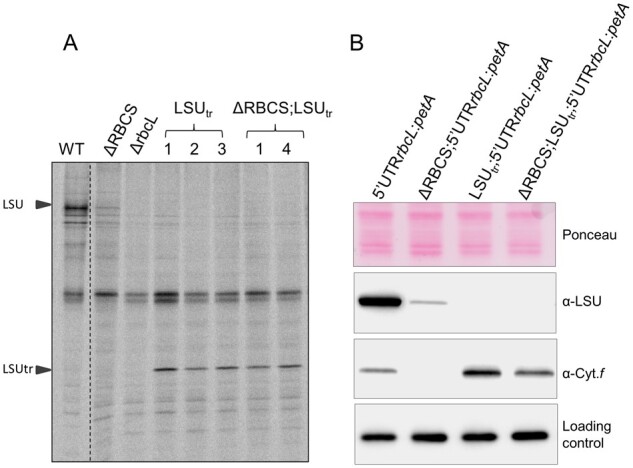
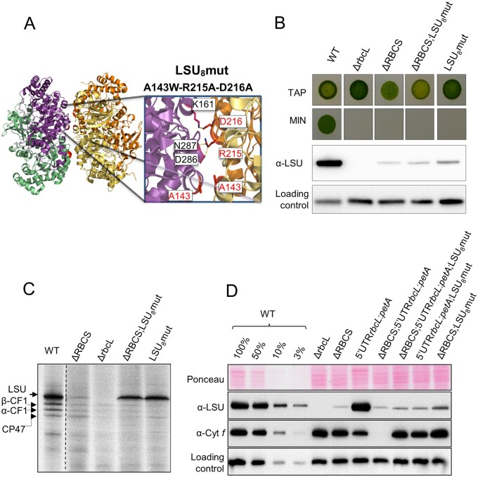
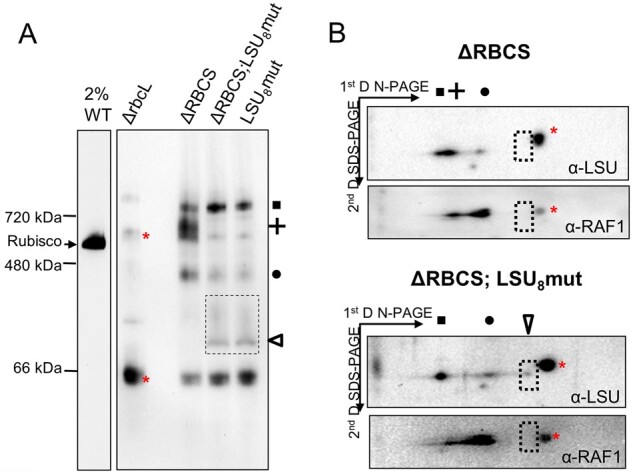
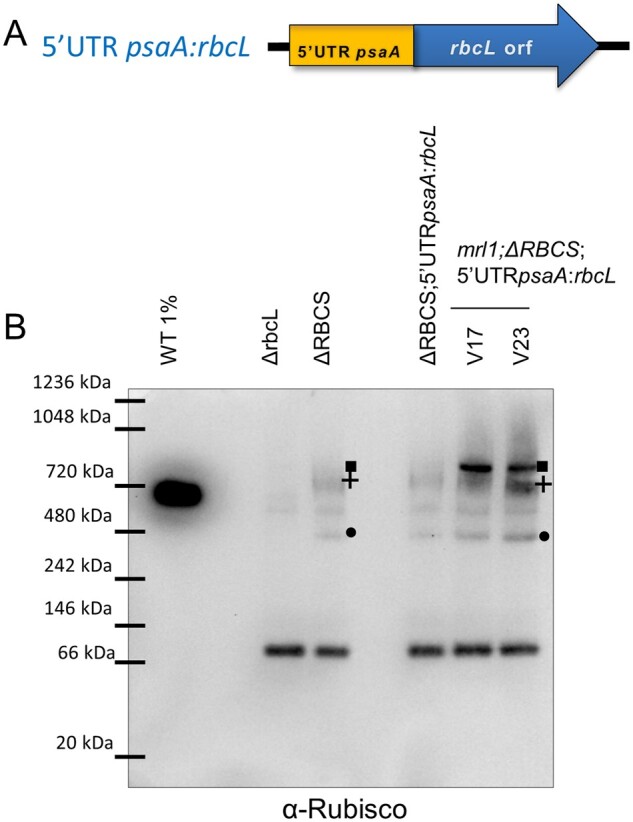
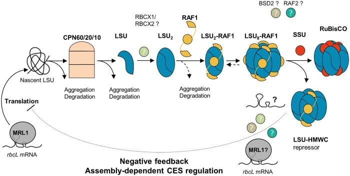
Ribulose 1,5-bisphosphate carboxylase/oxygenase (Rubisco) is present in all photosynthetic organisms and is a key enzyme for photosynthesis-driven life on Earth. Its most prominent form is a hetero-oligomer in which small subunits (SSU) stabilize the core of the enzyme built from large subunits (LSU), yielding, after a chaperone-assisted multistep assembly process, an LSU8SSU8 hexadecameric holoenzyme. Here we use Chlamydomonas reinhardtii and a combination of site-directed mutants to dissect the multistep biogenesis pathway of Rubisco in vivo. We identify assembly intermediates, in two of which LSU are associated with the RAF1 chaperone. Using genetic and biochemical approaches we further unravel a major regulation process during Rubisco biogenesis, in which LSU translation is controlled by its ability to assemble with the SSU, via the mechanism of control by epistasy of synthesis (CES). Altogether this leads us to propose a model whereby the last assembly intermediate, an LSU8-RAF1 complex, provides the platform for SSU binding to form the Rubisco enzyme, and when SSU is not available, converts to a key regulatory form that exerts negative feedback on the initiation of LSU translation.


Figures

Figure 1
Figure 1
LSU accumulation, synthesis rate, and stability in the absence of its assembly partner. (A) Immunoblot showing protein accumulation of Rubisco subunits in the ΔRBCS strain, using an antibody directed against whole Rubisco holoenzyme. PsaD accumulation, revealed with a specific antibody, is shown as a loading control. (B) 14C pulse labeling experiment showing the synthesis rate of LSU in the ΔRBCS strain as compared to the WT in upper panel (positions of LSU as well as ATPase α and β subunits and PSII CP43 subunit are indicated by arrows). (C) mRNA accumulation in the same strains as in B, as probed by hybridization with rbcL and RBCS probes, and psaB and CBLP probes used as loading controls. In both panels, the ΔrbcL strain exhibiting a deletion of the rbcL gene is used as a negative control. (D) Unassembled LSU stability assayed by immunochase over 4h after chloroplast synthesis arrest by chloramphenicol (CAP) addition. LSU is detected with the anti-Rubisco antibody, cyt f is used as a loading control.
Figure 2
Figure 2
Swapping rbcL 5′ UTR regulatory sequence impairs the CES regulation. (A) Upper panel: Photosynthetic growth phenotypes of 5′UTRpsaA:rbcL strains defective or not for Rubisco SSU, and accumulation of the corresponding Rubisco subunits tested by western blot analysis. Lower panel: In ΔRBCS;5′UTRpsaA:rbcL, LSU is accumulating to higher levels than in ΔRBCS. TAP stands for TAP medium, MIN is an acetate-free, phototrophy-selective medium. (B) 14C pulse labeling experiment showing LSU synthesis rate in 5′UTRpsaA:rbcL-background with and without small subunit compared with wild-type, ΔrbcL and ΔRBCS strains. The dashed line marks the position where two irrelevant lanes were removed.
Figure 3
Figure 3
Expression of cyt f is inhibited in the absence of Rubisco small subunit. Immunoblot using antibodies directed against the proteins depicted at the left, showing Rubisco and cyt f accumulation levels in the wild-type, ΔRBCS, ΔrbcL, and 5′UTRrbcL:petA strains with and without SSU. PsaD accumulation is shown as a loading control.
Figure 4
Figure 4
CES regulation does not occur in the absence of LSU accumulation. (A)14C labeling experiment showing synthesis rates of chloroplast proteins in WT, ΔrbcL, ΔRBCS, LSUtr transformants (1-3), and ΔRBCS;LSUtr (1 and 4) strains. Migration of full-length and truncated LSU is indicated on the left. The dashed line marks the position where two irrelevant lanes were removed. (B) Immunoblot depicting LSU and cyt f accumulation in representative transformants carrying both the 5′UTRrbcL:petA reporter gene and a truncation within the rbcL gene, associated or not to the ΔRBCS mutation, in comparison to the wild-type and ΔRBCS;5′UTRrbcL:petA strains. Ponceau stain and PsaD accumulation are shown as loading controls.
Figure 5
Figure 5
LSU assembly intermediates accumulate in the SSU-lacking strain. (A) Immunoblot with the antibody directed against Rubisco after native PAGE analysis of soluble protein extracts from WT (diluted to 2% as not to obscure the gel), ΔrbcL, and ΔRBCS strains. The migration of native molecular weight markers is indicated on the left. The position of Rubisco holoenzyme, as deduced from the WT signal, is indicated as well. Three LSU-specific complexes are observed in the SSU-lacking strain (depicted by a square, cross, and circle). (B) Analysis of the second dimension on SDS-PAGE gel by immunodetection of proteins putatively associated to LSU complexes in ΔRBCS strain (depicted by a square, cross, and circle as in (A), using anti-LSU, anti-CPN60, and anti-RAF1 antibodies. Dashed lines are drawn to help with the alignment. Red asterisks mark cross-contaminating signals of the anti-LSU antibody. (C) Immunochase in the ΔRBCS strain to follow the stability of the three LSU-oligomers detected in (A) (same symbols used) using a Rubisco antibody after native PAGE analysis as performed in (A). CAP, a chloroplast synthesis inhibitor, was added in the culture at the initial time point.
Figure 6
Figure 6
RAF1 and LSU interact in Chlamydomonas. (A) Immunoblots showing RAF1 and LSU co-immunoprecipitation. A similar fraction of the input, unbound (UB), or bead-extracted (E) fraction from the immunoprecipitation of soluble extracts from either the ΔRBCS or ΔRBCS-RAF1:Strep-TG strains was separated on SDS-PAGE gel, together with a molecular weight ladder (L). The line separates non-contiguous lanes of the same gel with the same exposure. RAF1 and LSU were detected by immunoblots using specific antibodies. The anti-RAF1 antibody recognizes both the endogenous (lower band) and the overexpressed epitope-tagged RAF1 (upper band), which could be separated by this gel system. The anti-Rubisco antibody recognizes LSU as well as an unrelated cross-reacting band marked by a red asterisk. LSU is specifically pulled-down by coimmunoprecipitation of the strep-tagged RAF1 protein. We note that not all LSU is pulled down, which could reveal the LSU fraction not associated to RAF1. (B) Strep-tagged RAF1 is associated to LSU LMW and HMW complexes, as shown by immunoblot using the RAF1 antibody and Rubisco antibody sequentially after separation of soluble proteins from the ΔRBCS-RAF1:Strep-TG and ΔRBCS on a 4–16% native gel followed by a second dimension in denaturating condition (10% SDS-PAGE, 8M urea gel). LSU complexes (depicted by a square, cross, and circle as in Figure 5A) in the ΔRBCS strain or ΔRBCS-RAF1:Strep-TG are shown on the top. Migration of the Strep-tagged or native RAF1 and of LSU is indicated on the left. The remaining observed signals come from cross-reactions with the antibodies. Note that here RAF1-related signals were left saturated, in order to properly see LSU-related signals.
Figure 7
Figure 7
RAF1 oligomerization state in Rubisco mutants versus WT. (A) Immunoblot showing similar RAF1 content in rbcL or RBCS deletion mutants (ΔrbcL and ΔRBCS strains), and in WT, using antibodies directed against RAF1, Rubisco, and PsaD, as a loading control. Note that Rubisco accumulation was probed from a distinct membrane part obtained after the transfer of duplicated samples on the same gel. (B) Immunoblot of a 1D native PAGE of soluble extracts from ΔrbcL, ΔRBCS, and WT using RAF1 (left and middle panels) or Rubisco antibody (right panel), showing that RAF1 accumulates as an oligomer in the absence of LSU. RAF1-LSU complexes are indicated using the same symbols as in Figure 5. Note that the RAF1-LSU HMW complex found in the ΔRBCS is no longer detected in a WT background, whereas an additional low abundant RAF1 complex, indicated by a black star, is found. Red asterisks indicate antibody cross-reacting bands. (The left panel is a distinct experiment from the middle and right panels, which were separated on the same gel).
Figure 8
Figure 8
LSU2 mutations alter LSU accumulation and CES regulation. (A) Close-up of the mutated residues in LSU2mut strain in LSU structure. C. reinhardtii LSU dimer structure is shown in cartoon, as extracted from Rubisco structure (PDB: 1IR2). The two LSU subunits forming the dimer are represented in green and magenta. Subunits are maintained by two inter-subunits salt bridges between E109 and R253, and E110 and R213 residues. Residues mutated in LSU2mut (E109A and R253A) are highlighted in red. The figure was generated using the PyMol program (Schrödinger-LLC). (B) Impairment in Rubisco accumulation is revealed by the absence of phototrophic growth in the LSU2mut and ΔRBCS;LSU2mut strains as probed by spot tests on acetate-free minimal media (MIN). Growth on TAP is shown as a control. The corresponding soluble LSU accumulation detected by immunoblot is shown together with PsaD accumulation as loading control. (C) LSU synthesis rate in LSU2mut and ΔRBCS;LSU2mut measured by short 14C pulse labeling experiment and compared to WT. Note that in the 12–18% acrylamide-8M urea gel system, the mutated LSU undergoes a change in its migration pattern compared to native LSU. (D) Immunoblot with the Rubisco antibody after CN-PAGE analysis of soluble protein fractions of WT (note the dilution), ΔrbcL, ΔRBCS, LSU2mut and ΔRBCS;LSU2mut strains. A dashed line marks the position where two irrelevant lanes were removed. The position of the LSU-complexes observed in ΔRBCS is indicated at the right using the same symbols as in Figure 5 (square, cross, and circle).
Figure 9
Figure 9
Disruption of LSU oligomerization alters LSU CES regulation. (A) Close-up of the mutated residues in the LSU8mut strain in LSU structure. Two LSU dimers facing each other are shown, as extracted from C. reinhardtii Rubisco structure (PDB: 1IR2). LSU subunits from the first and second depicted dimers are shown respectively in green and magenta, and in orange and yellow. The dimer to dimer interaction is stabilized by hydrogen bonds between the R215 and D286-N287 residues, and by a salt bridge involving the D216 and K161 residues, which are represented on the cartoon. The distance between the two dimers is the shortest at the A143 residues facing each other. Residues mutated in LSU8mut (ARD) are highlighted in red. The figure was generated using the PyMol program (Schrödinger-LLC). (B) Impairment in Rubisco accumulation is revealed by the absence of phototrophic growth in the LSU8mut and ΔRBCS; LSU8mut strains as probed by spot tests on acetate-free minimal media (MIN). Growth on TAP is provided as a control. Control strains WT, ΔrbcL, and ΔRBCS come from the same cultures as the ones used in Figure 7A. The corresponding soluble LSU accumulation detected by immunoblot is shown. (C) LSU synthesis rate in LSU8mut and ΔRBCS;LSU8mut measured by short 14C pulse labeling experiment and compared to ΔRBCS and WT. The dashed line marks the position of two irrelevant lanes, which were removed. (D) Immunoblot showing LSU and cyt f accumulation levels in the wild-type (WT), ΔRBCS, ΔrbcL, LSU8mut, and ΔRBCS;LSU8mut strains and in those latter three genetic contexts combined with the 5′UTRrbcL:petA reporter gene background. PsaD accumulation is provided as a loading control.
Figure 10
Figure 10
Alteration of CES regulation is concurrent with the disappearance of the LSU8-RAF1 oligomer. (A) Immunoblot with the Rubisco antibody after CN-PAGE analysis of soluble protein fractions of WT (note the dilution), ΔrbcL, ΔRBCS;LSU8mut, and ΔRBCS;LSU8mut strains. The position of the LSU-complexes observed in ΔRBCS is indicated at the right using the same symbols as in Figure 5 (square, cross, and circle). The empty triangle and dashed box indicate the somewhat diffuse band attributed to LSU dimer. (B) Immunoblot after CN-PAGE analysis (4–16%) of soluble protein fractions from ΔRBCS (top) and ΔRBCS;LSU8mut (bottom), followed by a second dimension run on a 13% SDS-PAGE gel using the anti-Rubisco and anti-RAF1 antibodies sequentially. The Rubisco antibody was stripped before rehybridization with the anti-RAF1 antibody, however a cross-reacting signal labeled with a red cross could not be completely stripped off. The position of the LSU-complexes observed in ΔRBCS and ΔRBCS;LSU8mut are indicated on top of the gels using the same symbols as in A (square: LSU-CPN60, cross: LSU8-RAF1 and circle: LSU2-RAF1). The empty triangle denotes the band observed in RBCS;LSU8mut attributed to RAF1-free LSU dimers. No corresponding signal can be detected at this position (dashed rectangle) with the RAF1 antibody in the ΔRBCS strain.
Figure 11
Figure 11
The LSU-HMW complex is not affected by MRL1 absence in the mrl1;ΔRBCS;5′UTRpsaA:rbcL strain. (A) Scheme of the 5′UTR psaA:rbcL chimeric construct. (B) Native immunoblot using the Rubisco antibody in order to follow the migration pattern of the LSU-HMW repressor complex using soluble proteins extracted from strains expressing LSU from the 5′UTRpsaA:rbcL chloroplast transgene in an RBCS mutant, in a MRL1 WT background (ΔRBCS; 5′UTRpsaA:rbcL) or mutant background (mrl1; ΔRBCS; 5′UTRpsaA:rbcL V17 and V23). WT diluted extract, as well as extracts from the ΔrbcL and ΔRBCS strains, were included as controls. LSU oligomers are depicted by the same symbols used in Figure 5.
Figure 12
Figure 12
Model of Rubisco biogenesis pathway and CES regulation. The rbcL mRNA, stabilized by the binding of the MRL1 PPR-protein to its 5′UTR region, can be translated. Nascent LSU is recruited by the chloroplast folding machinery. LSU propeptide is subsequently folded in the CPN60/20/10 chaperonin complex. The released LSU dimerizes, maybe with help of RBCX, and recruits RAF1 which is required for LSU2 stabilization. LSU2-RAF1 unit oligomerizes further to form Rubisco catalytic core. RAF1 is finally substituted by the SSU to form the complete holoenzyme. In the SSU-limiting context, the LSU-RAF1 HMWC is converting to a repressor of rbcL translation (CES process) preventing LSU wasteful production, by binding either directly rbcL mRNA or other factors, thereby displacing some RAF1 oligomers. Many aspects of this model remain unclear such as the identity of the proteins/RNA in the LSU regulator complex, or the exact role of the other Rubisco assembly chaperones such as RBCX1/2 and RAF2, or the presence of a functional homolog of the plant BSD2 factor in algae, which remains debated.
None

Similar articles

Cited by

References

    1. Aigner H, Wilson RH, Bracher A, Calisse L, Bhat JY, Hartl FU, Hayer-Hartl M (2017) Plant RuBisCo assembly in E. coli with five chloroplast chaperones including BSD2. Science 358: 1272–1278 - PubMed
    1. Andersson I, Backlund A (2008) Structure and function of Rubisco. Plant Physiol Biochem 46: 275–291 - PubMed
    1. Bar-On YM, Milo R (2019) The global mass and average rate of rubisco. Proc Natl Acad Sci USA 116: 4738–4743 - PMC - PubMed
    1. Barkan A (1993) Nuclear mutants of maize with defects in chloroplast polysome assembly have altered chloroplast RNA metabolism. Plant Cell 5: 389–402 - PMC - PubMed
    1. Barkan A, Small I (2014) Pentatricopeptide repeat proteins in plants. Annu Rev Plant Biol 65: 415–442 - PubMed

Publication types

MeSH terms

Substances