. 2021 Jun;18(3):411-427.
doi: 10.1007/s13770-021-00330-7. Epub 2021 Feb 24.

Growth and Differentiation of Circulating Stem Cells After Extensive Ex Vivo Expansion

Affiliations

Growth and Differentiation of Circulating Stem Cells After Extensive Ex Vivo Expansion

Silvia Barbon et al. Tissue Eng Regen Med. 2021 Jun.

Abstract

Background: Stem cell therapy is gaining momentum as an effective treatment strategy for degenerative diseases. Adult stem cells isolated from various sources (i.e., cord blood, bone marrow, adipose tissue) are being considered as a realistic option due to their well-documented therapeutic potentials. Our previous studies standardized a method to isolate circulating multipotent cells (CMCs) that are able to sustain long term in vitro culture and differentiate towards mesodermal lineages.

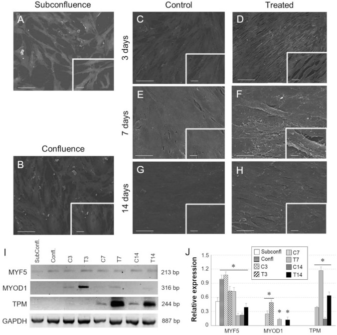
Methods: In this work, long-term cultures of CMCs were stimulated to study in vitro neuronal and myogenic differentiation. After induction, cells were analysed at different time points. Morphological studies were performed by scanning electron microscopy and specific neuronal and myogenic marker expression were evaluated using RT-PCR, flow cytometry and western blot. For myogenic plasticity study, CMCs were transplanted into in vivo model of chemically-induced muscle damage.

Results: After neurogenic induction, CMCs showed characteristic dendrite-like morphology and expressed specific neuronal markers both at mRNA and protein level. The calcium flux activity of CMCs under stimulation with potassium chloride and the secretion of noradrenalin confirmed their ability to acquire a functional phenotype. In parallel, the myogenic potential of CMCs was confirmed by their ability to form syncytium-like structures in vitro and express myogenic markers both at early and late phases of differentiation. Interestingly, in a rat model of bupivacaine-induced muscle damage, CMCs integrated within the host tissue taking part in tissue repair.

Conclusion: Overall, collected data demonstrated long-term cultured CMCs retain proliferative and differentiative potentials suggesting to be a good candidate for cell therapy.

Keywords: Circulating stem cells; Degenerative diseases; Myogenesis; Neurogenesis; Regenerative medicine.


Conflict of interest statement

The Authors declare that there is no conflict of interest regarding the publication of this article.

Figures

Fig. 1
Fig. 1
In vitro characterization of CMCs. A Morphological analysis by SEM. Scale bar: 300 µm. B Population doubling study. C Karyotype analysis. D Immunophenotyping by flow cytometry. E Pluripotency gene expression profile identification by qPCR. F Adipogenic (Oil Red O) and G osteogenic (Von Kossa) differentiation responses. Scale bar: F 10 µm; G 50 µm. H SPARC gene expression detected by RT-PCR (top) and densitometry band quantification (bottom) in CMCs after 20 days of chondrogenic induction (treated cells) in comparison with the control (untreated cells). (*p < 0.05)
Fig. 2
Fig. 2
A–F Morphological study by SEM of CMCs cultured in NeuroBasal Medium (A, B), treated with EGF (20 ng/mL) and bFGF (10 ng/mL) for 7 days (C, D) and then with Retinoid Acid (RA) (0.5 µM) and NGF (20 ng/mL) up to 14 days (E, F). Scale bar: A, C, E 50 µm; B, D, F 10 µm. G OneStep RT-PCR study of neuronal marker expression of CMCs grown in proliferation medium (Undiff), control cells (C7) and cells treated for 7 and 14 days (T7, T14) with inductive factors. In parallel, the expression of the housekeeping gene GAPDH was detected in all samples. H Relative quantification of electrophoresis gel bands by densitometric analysis. (*p ≤ 0.05 vs. undifferentiated sample)
Fig. 3
Fig. 3
A Flow cytometrical analysis of TBB3, MAP2, TH on induced (T14) CMCs (coloured profile) compared to cells cultured in αMEM, NBM, and NBM supplemented with neurogenic factors for 14 days (black profile for all). Indirect staining with FITC-conjugated secondary antibodies was used. For each marker, data were expressed as % positives ± SD of T14 versus C14. B Western Blot analysis on undifferentiated (Undiff), C7, T7 and T14 CMCs using 10 g protein extract and chemiluminescence detection. C Densitometric quantification of Western Blot bands. (*p ≤ 0.05 vs undifferentiated sample). D Immunofluorescence of neuronal lineage markers on induced CMCs (T14) compared to undifferentiated samples (Undiff). Cells were indirectly labeled using FITC-conjugated secondary antibodies and images were acquired with the Leica TCS SP5 confocal microscope. Scale bar: 50 µm
Fig. 4
Fig. 4
A Noradrenaline release by HPLC in CMC cultures after neurogenic induction. B Emission spectrum of the fluorescent calcium indicator Indo1 AM (5 μM) after incubation (30 min, 37 °C) with differentiated CMC cultures (C14, T14) previously treated with KCl 56 mM
Fig. 5
Fig. 5
A–H Morphological study by SEM of CMC cultures at subconfluent (subconfl) state, at 100% confluence (Confl) and ± treatment with myogenic inductive medium for 3, 7 and 14 days. Scale bar: AD 100 µm; EH 50 µm. I Gene expression analysis by One step RT-PCR of myogenic markers in differentiated CMC cultures. GAPDH was taken as housekeeping gene. J Densitometric quantification of Western Blot bands by ImageJ software. (*p ≤ 0.05 vs. cultures at subconfluent state)
Fig. 6
Fig. 6
AE Protein expression analysis by Western Blot flow cytometry and immunofluorescence of myogenic markers in CMCs differentiated towards myogenic lineage. B Quantification of WB bands by densitometry analysis. (*p ≤ 0.05 vs. cells at subconfluent state). C, D Data are expressed as percentage of positive induced cells compared to undifferentiated control cells. (Subconfl: cultures at a subconfluent state; Confl: cells at 100% confluence). E Scale bar: 50 µm
Fig. 7
Fig. 7
A In vivo experimental plan. B–G Immunofluorescence of vimentin in bupivacaine-damaged tibialis anterior muscle after 7 (B, D, F) and 14 (C, E, G) days from injection of Q Dot labeled CMCs. Scale bar: BG 100 µm; B–G 25 µm

References

    1. Ramaswami M, Taylor JP, Parker R. Altered ribostasis: RNA-protein granules in degenerative disorders. Cell. 2013;154:727–736. doi: 10.1016/j.cell.2013.07.038. - DOI - PMC - PubMed
    1. Paolini Paoletti F, Gaetani L, Parnetti L. Molecular profiling in Parkinsonian syndromes: CSF biomarkers. Clin Chim Acta. 2020;506:55–66. doi: 10.1016/j.cca.2020.03.002. - DOI - PubMed
    1. Castillo X, Castro-Obregón S, Gutiérrez-Becker B, Gutiérrez-Ospina G, Karalis N, Khalil AA, et al. Re-thinking the etiological framework of neurodegeneration. Front Neurosci. 2019;13:728. doi: 10.3389/fnins.2019.00728. - DOI - PMC - PubMed
    1. Dugger BN, Dickson DW. Pathology of neurodegenerative diseases. Cold Spring Harb Perspect Biol. 2017;9:a028035. doi: 10.1101/cshperspect.a028035. - DOI - PMC - PubMed
    1. Bond AM, Ming GL, Song H. Adult mammalian neural stem cells and neurogenesis: five decades later. Cell Stem Cell. 2015;17:385–395. doi: 10.1016/j.stem.2015.09.003. - DOI - PMC - PubMed

Publication types

LinkOut - more resources