. 2021 Jun;53(6):1766-1790.
doi: 10.1002/jmri.27542. Epub 2021 Feb 24.

Systematic Review of Magnetic Resonance Lymphangiography From a Technical Perspective

Affiliations

Systematic Review of Magnetic Resonance Lymphangiography From a Technical Perspective

Michael Mills et al. J Magn Reson Imaging. 2021 Jun.

Abstract

Background: Clinical examination and lymphoscintigraphy are the current standard for investigating lymphatic function. Magnetic resonance imaging (MRI) facilitates three-dimensional (3D), nonionizing imaging of the lymphatic vasculature, including functional assessments of lymphatic flow, and may improve diagnosis and treatment planning in disease states such as lymphedema.

Purpose: To summarize the role of MRI as a noninvasive technique to assess lymphatic drainage and highlight areas in need of further study.

Study type: Systematic review.

Population: In October 2019, a systematic literature search (PubMed) was performed to identify articles on magnetic resonance lymphangiography (MRL).

Field strength/sequence: No field strength or sequence restrictions.

Assessment: Article quality assessment was conducted using a bespoke protocol, designed with heavy reliance on the National Institutes of Health quality assessment tool for case series studies and Downs and Blacks quality checklist for health care intervention studies.

Statistical tests: The results of the original research articles are summarized.

Results: From 612 identified articles, 43 articles were included and their protocols and results summarized. Field strength was 1.5 or 3.0 T in all studies, with 25/43 (58%) employing 3.0 T imaging. Most commonly, imaging of the peripheries, upper and lower limbs including the pelvis (32/43, 74%), and the trunk (10/43, 23%) is performed, including two studies covering both regions. Imaging protocols were heterogenous; however, T2 -weighted and contrast-enhanced T1 -weighted images are routinely acquired and demonstrate the lymphatic vasculature. Edema, vessel, quantity and morphology, and contrast uptake characteristics are commonly reported indicators of lymphatic dysfunction.

Data conclusion: MRL is uniquely placed to yield large field of view, qualitative and quantitative, 3D imaging of the lymphatic vasculature. Despite study heterogeneity, consensus is emerging regarding MRL protocol design. MRL has the potential to dramatically improve understanding of the lymphatics and detect disease, but further optimization, and research into the influence of study protocol differences, is required before this is fully realized.

Level of evidence: 2 TECHNICAL EFFICACY: Stage 2.

Keywords: Magnetic resonance lymphangiography; lymphatics; lymphedema; lymphography; review.


Figures

Figure 1
Figure 1
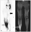
Lymphoscintigram (a) and magnetic resonance lymphangiogram (b) acquired in the lower limbs of a participant with lymphedema of the right lower limb. MRI was acquired after contrast injection in the affected limb with a contrast-enhanced 3D T1-weighted gradient echo sequence with TR/TE = 4.13/1.47 msec, flip angle = 25°, reconstructed voxel size = 0.80 × 0.80 × 0.80 mm. Both modalities show regions of dermal reflex (open arrows). The lymphoscintigram also shows a normal appearing main lymphatic pathway leading to the inguinal lymph nodes in the unaffected (left) limb (filled arrow). Reproduced from Weiss et al., with permission.
Figure 2
Figure 2
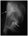
Lower limb indocyanine green (ICG) fluorescence image, showing the lateral aspect of the shin, in a participant with unilateral lower limb lymphedema acquired by St George’s Lymphovascular Research Group. ICG binds to proteins such as albumin making imaging specific to the lymphatics. This image was produced via laser excitation of the ICG after intradermal injection between the digital webspaces, and subsequent detection of the fluorescence by a CCD detector. High spatial resolution allows identification of individual superficial lymphatic vessel (solid arrow); however, emissions from deeper lying structures are quickly attenuated. In the unaffected individual, fairly linear vessel pathways flowing distally to proximally, and following known anatomical pathways, should be observed. In an affected state, an abnormal drainage pattern is evident such as no flow, medial to lateral (or vice versa) flow, and dermal rerouting (dashed arrow). Image “Lower limb ICG in unilateral lymphedema” shared by St George’s Lymphovascular Research Group under the CC BY-SA-4.0 International license (https://creativecommons.org/licenses/by-sa/4.0/). https://commons.wikimedia.org/wiki/File:Lower_Limb_ICG_in_unilateral_lymphoedema.tif.
Figure 3
Figure 3
Study selection flow chart. PubMed revealed 609 English language sources after a search for lymphatic vessel magnetic resonance imaging. After vetting and quality assessment, a total of 43 articles were included in this review, the majority of which report imaging in the limbs and/or pelvis (collectively labeled the “peripheries”). Note that some studies cover both the torso and the limbs and so are counted twice. One study, performing peripheral MRL and a single case of torso MRL, was included for review with the single torso case excluded.
Figure 4
Figure 4
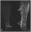
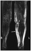
Maximum intensity projected T2-weighted noncontrast MRL image of a participant with unilateral lymphedema of the left leg. TR/TE = 4000/884 msec, flip angle = 90°, voxel size = 0.8 × 1.4 mm, acquired with a driven equilibrium pulse. Many tortuous vessel-like structures are seen in the left leg (solid arrows), with signal intense areas of fluid accumulation seen by the left ankle (dashed arrows). High signal structures are also observed at the right ankle (diamond headed arrow). The high signal in the vessel-like structures seen in the left limb may be due to vessel dilation and/or fluid stasis, both of which can occur as a result of pathology. Reproduced from Arrive et al., with permission.
Figure 5
Figure 5
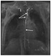
Thoracic duct MRL of a participant with bilateral upper and lower limb lymphedema acquired with a contrast-enhanced T1-weighted SPGR by St George’s Lymphovascular Research Group. TR/TE = 5.2 / 1.8 msec, flip angle = 30°, reconstructed voxel size = 0.75 × 0.75 × 1.50 mm. This MIP clearly displays contrast draining through a single smooth channeled thoracic duct (solid arrow), which appears to bifurcate and drain bilaterally (dashed arrows). Image “Thoracic duct MRL in lymphedema” shared by St George’s Lymphovascular Research Group under the CC BY-SA-4.0 International license (https://creativecommons.org/licenses/by-sa/4.0/). https://commons.wikimedia.org/wiki/File:Thoracic_duct_MRL_in_lymphoedema.tif.
Figure 6
Figure 6
T2-weighted TSE image of a participant with lower limb lymphedema in the left limb demonstrating a clear honeycomb pattern of the subcutaneous tissue (arrow). Acquired with TR/TE = 2870/797 msec, voxel size = 1.1 × 1.0 × 1.0 mm. Reproduced from Cellina et al., with permission.
Figure 7
Figure 7
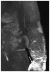
Contrast-enhanced image of the left arm of an individual with lymphedema showing a region of dermal backflow, the rerouting of lymph to the dermal lymphatics. Acquired with a fat suppressed SPGR, TR/TE = 3.5/1.3 msec, flip angle = 14.9°, voxel size = 1.0 × 1.4 × 1.2 mm. Reproduced from Bae et al., with permission.
Figure 8
Figure 8
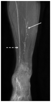
Lower limb MRL of a healthy participant imaged with a fat suppressed contrast-enhanced T1 weighted SPGR by St George’s Lymphovascular Research Group. TR/TE = 3.6 / 1.6 msec, flip angle = 12°, reconstructed voxel size = 0.75 × 0.75 × 0.75 mm. This MIP demonstrates thin, discontinuous appearing, lymphatic vessels (solid arrow), as well as larger venous structures (dashed arrow). Image “Lower limb MRL in healthy participant” shared by St George’s Lymphovascular Research Group under the CC BY-SA-4.0 International license (https://creativecommons.org/licenses/by-sa/4.0/). https://commons.wikimedia.org/wiki/File:Lower_Limb_MRL_in_healthy_participant.tif.
Figure 9
Figure 9
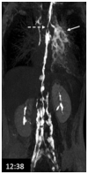
Lymphatic leakage (solid arrow) and thoracic duct narrowing (dashed arrow) identified 12 minutes into imaging of a patient with recurrent chylothorax. Acquired with a fat suppressed SPGR, TR/TE = 4.0/1.9 msec, flip angle = 10°, voxel size = 1.0 × 1.4 × 1.2 mm. Reproduced from Krishnamurthy et al., with permission.
Figure 10
Figure 10
Time of flight (TOF) image in the head of a healthy volunteer showing signal in the meningeal lymphatics (arrow) and low signal in the superior sagittal sinus, SSS (arrow head). Image produced with TR/TE = 30/4.49 msec, flip angle = 10°, voxel size = 0.31 × 0.31 × 1.5 mm, and subtracting images acquired with saturation bands anterior and posterior to the SSS from those acquired with a saturation band only anterior to the SSS. Reproduced from Kuo et al., with permission.

References

    1. Sleigh BC, Manna B. Lymphedema. StatPearls Publishing; Treasure Island, FL: 2020. Available at: https://www.ncbi.nlm.nih.gov/books/NBK537239/ - PubMed
    1. Földi M, Földi E. Földi’s textbook of lymphology for physicians and lymphoedmema therapists. 3rd ed. Urban & Fischer; München: 2012.
    1. Schulze H, Nacke M, Gutenbrunner C, Hadamitzky C. Worldwide assessment of healthcare personnel dealing with lymphoedema. Health Econ Rev. 2018;8:10. - PMC - PubMed
    1. The International Society of Lymphology. The diagnosis and treatment of peripheral lymphedema: 2020 consensus document of the International Society of Lymphology. Lymphology. 2020;53:3–19. - PubMed
    1. Weiss M, Burgard C, Baumeister R, et al. Magnetic resonance imaging versus lymphoscintigraphy for the assessment of focal lymphatic transport disorders of the lower limb. Nuklearmedizin. 2014;53:190–196. - PubMed

Publication types