. 2021 Apr 15;32(8):675-689.
doi: 10.1091/mbc.E20-03-0190. Epub 2021 Feb 24.

Rpgrip1l controls ciliary gating by ensuring the proper amount of Cep290 at the vertebrate transition zone

Affiliations

Rpgrip1l controls ciliary gating by ensuring the proper amount of Cep290 at the vertebrate transition zone

Antonia Wiegering et al. Mol Biol Cell. .

Abstract

A range of severe human diseases called ciliopathies is caused by the dysfunction of primary cilia. Primary cilia are cytoplasmic protrusions consisting of the basal body (BB), the axoneme, and the transition zone (TZ). The BB is a modified mother centriole from which the axoneme, the microtubule-based ciliary scaffold, is formed. At the proximal end of the axoneme, the TZ functions as the ciliary gate governing ciliary protein entry and exit. Since ciliopathies often develop due to mutations in genes encoding proteins that localize to the TZ, the understanding of the mechanisms underlying TZ function is of eminent importance. Here, we show that the ciliopathy protein Rpgrip1l governs ciliary gating by ensuring the proper amount of Cep290 at the vertebrate TZ. Further, we identified the flavonoid eupatilin as a potential agent to tackle ciliopathies caused by mutations in RPGRIP1L as it rescues ciliary gating in the absence of Rpgrip1l.


Figures

FIGURE 1:
FIGURE 1:
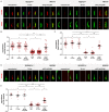
In mouse cells, ciliary gating is disturbed by the loss of Rpgrip1l but not by the loss of Nphp4. (A) Immunofluorescence on MEFs obtained from WT (n = 5) and Rpgrip1l–/– (n = 5) embryos. At least 10 cilia per embryo were used for quantifications (Σ(WT) = 62 cilia, Σ(Rpgrip1l–/–) = 72 cilia). (B) Immunofluorescence on MEFs obtained from WT (Arl13b: n = 4; Sstr3: n = 3) and Nphp4–/– (Arl13b: n = 4; Sstr3: n = 3) embryos. At least 20 cilia per embryo were used for quantifications (Arl13b: Σ(WT) = 83 cilia, Σ(Nphp4–/–) = 171 cilia; Sstr3: Σ(WT) = 134 cilia, Σ(Nphp4–/–) = 92 cilia). (A, B) The ciliary axoneme is stained in green by acetylated α-tubulin, the BB is stained in blue by γ-tubulin. The scale bars represent a length of 0.5 µm. Data are shown as mean ± SEM. Asterisks denote statistical significance according unpaired t tests with Welch´s correction (***P < 0.001) (A: t (25) = 6.54, P < 0.0001; B: Arl13b: t (46) = 0.7483, P < 0.4581; Sstr3: t (80) = 1.807, P < 0.0745).
FIGURE 2:
FIGURE 2:
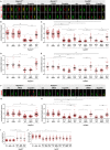
Loss of Cep290 but not of Nphp1 or Invs impairs ciliary gating in mouse cells. (A, B) Immunofluorescence on NIH3T3 cells. The ciliary axoneme is stained in green by acetylated α-tubulin. The scale bars represent a length of 0.5 µm. At least 20 cilia per clone were used for quantification. Data are shown as mean ± SEM. Asterisks denote statistical significance according to one-way ANOVA and Tukey HSD tests (***P < 0.001) (A: F (7,160) = 26.09, P < 0.0001; B: F (7,147) = 133.6, P < 0.0001).
FIGURE 3:
FIGURE 3:
Rescue of the Cep290 amount at the TZ of Rpgrip1l–/– NIH3T3 and RPGRIP1L–/– HEK293 cells restores the ciliary Arl13b and Sstr3 amount. (A–C) Rpgrip1l+/+ and Rpgrip1l–/– NIH3T3 cells were either untransfected or transfected with TF-Ctrl or pFlag-mCep290. (A) Immunofluorescence on Rpgrip1l+/+ and Rpgrip1l–/– (clone 10-61) NIH3T3 cells. The ciliary axoneme is stained in green by acetylated α-tubulin and Arl13b or Sstr3 is stained in red. The scale bars represent a length of 0.5 µm. (B, C) Normalized ciliary amount of Arl13b (B) and Sstr3 (C). At least 30 cilia per clone were used for quantification. Data are shown as mean ± SEM. Asterisks denote statistical significance according to one-way ANOVA and Tukey HSD tests (**P < 0.01; ***P < 0.001) (B: F (5, 400) = 146.8, P < 0.0001; C: F (5, 166) = 128.6, P < 0.0001). (D, E) RPGRIP1L+/+ and RPGRIP1L–/– HEK293 cells were either untransfected or transfected with eGFP-hCEP290 or TF-Ctrl. (D) Immunofluorescence on RPGRIP1L+/+ and RPGRIP1L–/– (clone 1–7) HEK293 cells. The ciliary axoneme is stained in green by acetylated α-tubulin and Arl13b is stained in red. The scale bars represent a length of 0.5 µm. (E) Normalized ciliary amount of Arl13b. At least 10 cilia (RPGRIP1L+/+HEK293 cells) or 20 cilia (RPGRIP1L–/– HEK293 cells) per clone were used for quantification. Data are shown as mean ± SEM. Asterisks denote statistical significance according to one-way ANOVA and Tukey HSD tests (***P < 0.001) (F (5, 104) = 142, P < 0.0001).
FIGURE 4:
FIGURE 4:
Eupatilin treatment rescues ciliary gating in the absence of Rpgrip1l. (A, B) Immunofluorescence on Rpgrip1l+/+, Rpgrip1l–/– (clone 10-61) and Cep290–/– NIH3T3 cells. The ciliary axoneme is stained in green by acetylated α-tubulin. Arl13b or Sstr3 is stained in red. The scale bars represent a length of 1 µm. (B–E) Normalized ciliary amount of Arl13b (B, C) and Sstr3 (D, E). At least 20 (D, E) or 30 cilia (B, C) per clone were used for quantification. The same quantification of WT serves as comparison to Rpgri1l-negative and Cep290-negative cells, respectively (B and C; D and E). Data are shown as mean ± SEM. Asterisks denote statistical significance according to one-way ANOVA and Tukey HSD tests (***P < 0.001) (B: F (5, 447) = 117.1, P < 0.0001; C: F (11, 690) = 163.4, P < 0.0001; D: F (5, 183) = 47.48, P < 0.0001; E: F (11, 393) = 35.27, P < 0.0001). (F) Immunofluorescence on Rpgrip1l+/+, Rpgrip1l–/– (clone 10-61) and Cep290–/– (clones 39-10, 39-51, 39-53) NIH3T3 cells. The ciliary axoneme is stained in green by acetylated α-tubulin. Nphp5 is stained in red. The scale bars represent a length of 1 µm. (G, H) Normalized ciliary amount of Nphp5. At least 40 cilia per clone were used for quantification. The same quantification of WT serves as comparison to Rpgrip1l-negative and Cep290-negative cells. Data are shown as mean ± SEM. Asterisks denote statistical significance according to one-way ANOVA and Tukey HSD tests (***P < 0.001) (G: F (5, 354) = 44.34, P < 0.0001; H: F (11, 638) = 64.13, P < 0.0001). (I, J) Ciliary length measurements. At least 30 cilia (I) or 50 cilia (J) per clone were used for quantification. The same quantification of the WT serves as comparison to Rpgrip1l-negative and Cep290-negative cells. Data are shown as mean ± SEM. Asterisks denote statistical significance according to one-way ANOVA and Tukey HSD tests (***P < 0.001) (I: F (5, 402) = 28.71, P < 0.0001; J: F (11, 986) = 36,73, P < 0.0001).
FIGURE 5:
FIGURE 5:
Graphical abstract of ciliary gating defects and their rescue in Rpgrip1l- and Cep290-negative NIH3T3 cells. Rpgrip1l controls ciliary gating via ensuring the proper amount of Cep290 at the TZ, which in turn regulates the ciliary amount of Nphp5. In the absence of Rpgrip1l, the ciliary amount of Cep290 and Nphp5 is reduced. This leads to ciliary gating defects indicated by lower ciliary Arl13b and Sstr3 levels. In Cep290-negative cells, the ciliary amount of Rpgrip1l is unaltered, whereas the ciliary amount of Nphp5 is reduced. This leads to down-regulation of ciliary gating in Cep290–/– cells. Restoration of the Cep290 amount in Rpgrip1l–/– cells via transfection of full-length Cep290 rescues ciliary gating defects. We propose that the rescue of ciliary gating in transfected Rpgrip1l–/–cells is mediated by a restored ciliary amount of Nphp5.

Similar articles

Cited by

References

    1. Andreu-Cervera A, Anselme I, Karam A, Laclef C, Catala M, Schneider-Maunoury S (2019). The ciliopathy gene ftm/rpgrip1l controls mouse forebrain patterning via region-specific modulation of hedgehog/gli signaling. J Neurosci 39, 2398–2415. - PMC - PubMed
    1. Assis L, Silva-Junior R, Dolce L, Alborghetti M, Honorato R, Nascimento A, Melo-Hanchuk T, Trindade D, Tonoli C, Santos C, et al. (2017). The molecular motor Myosin Va interacts with the cilia-centrosomal protein RPGRIP1L. Sci Rep 7, 43692. - PMC - PubMed
    1. Aubusson-Fleury A, Lemullois M, de Loubresse N, Laligné C, Cohen J, Rosnet O, Jerka-Dziadosz M, Beisson J, Koll F (2012). The conserved centrosomal protein FOR20 is required for assembly of the transition zone and basal body docking at the cell surface. J Cell Sci 125, 4395–4404. - PubMed
    1. Avasthi P, Marshall W (2012). Stages of ciliogenesis and regulation of ciliary length. Differentiation 83, 30–42. - PMC - PubMed
    1. Awata J, Takada S, Standley C, Lechtreck K, Bellvé K, Pazour G, Fogarty K, Witman G (2014). NPHP4 controls ciliary trafficking of membrane proteins and large soluble proteins at the transition zone. J Cell Sci 127, 4714–4727. - PMC - PubMed

Publication types

MeSH terms

LinkOut - more resources