

Comparative analysis of ACE2 protein expression in rodent, non-human primate, and human respiratory tract at baseline and after injury: A conundrum for COVID-19 pathogenesis
- PMID: 33626084
- PMCID: PMC7904186
- DOI: 10.1371/journal.pone.0247510
Comparative analysis of ACE2 protein expression in rodent, non-human primate, and human respiratory tract at baseline and after injury: A conundrum for COVID-19 pathogenesis
Abstract
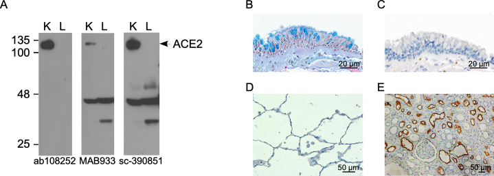
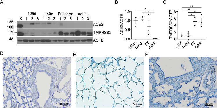
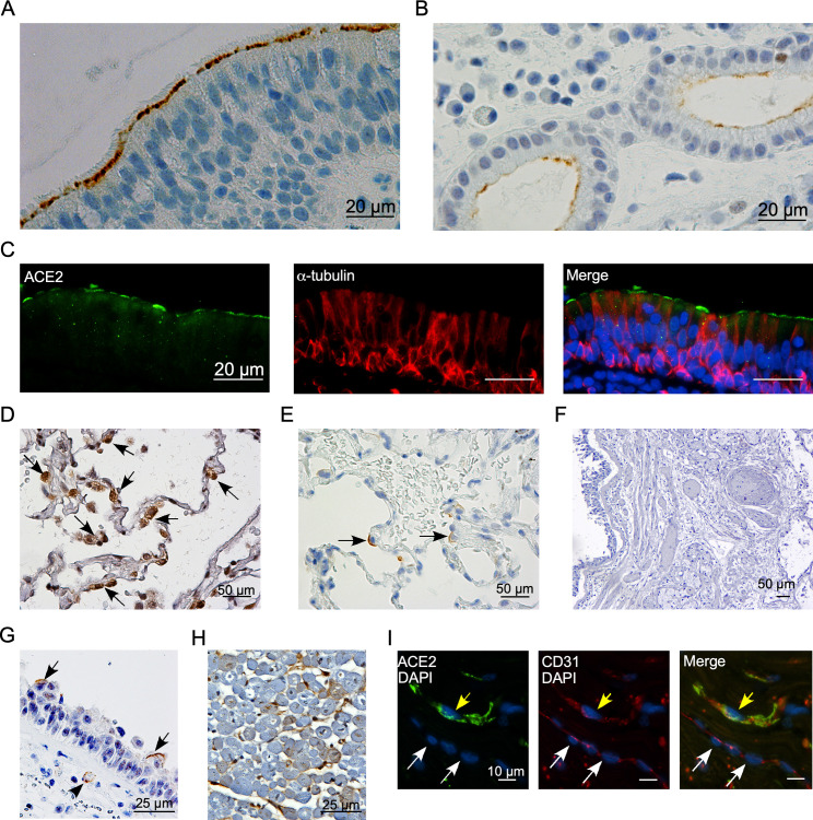
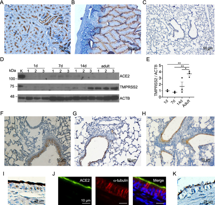
Angiotensin converting enzyme 2 (ACE2) is the putative functional receptor for severe acute respiratory syndrome coronavirus 2 (SARS-CoV-2). Current literature on the abundance and distribution of ACE2 protein in the human respiratory tract is controversial. We examined the effect of age and lung injury on ACE2 protein expression in rodent and non-human primate (NHP) models. We also examined ACE2 expression in human tissues with and without coronavirus disease 19 (COVID-19). ACE2 expression was detected at very low levels in preterm, but was absent in full-term and adult NHP lung homogenates. This pattern of ACE2 expression contrasted with that of transmembrane protease serine type 2 (TMPRSS2), which was significantly increased in full-term newborn and adult NHP lungs compared to preterm NHP lungs. ACE2 expression was not detected in NHP lungs with cigarette smoke-induced airway disease or bronchopulmonary dysplasia. Murine lungs lacked basal ACE2 immunoreactivity, but responded to hyperoxia, bacterial infection, and allergen exposure with new ACE2 expression in bronchial epithelial cells. In human specimens, robust ACE2 immunoreactivity was detected in ciliated epithelial cells in paranasal sinus specimens, while ACE2 expression was detected only in rare type 2 alveolar epithelial cells in control lungs. In autopsy specimens from patients with COVID-19 pneumonia, ACE2 was detected in rare ciliated epithelial and endothelial cells in the trachea, but not in the lung. There was robust expression of ACE2 expression in F344/N rat nasal mucosa and lung specimens, which authentically recapitulated the ACE2 expression pattern in human paranasal sinus specimens. Thus, ACE2 protein expression demonstrates a significant gradient between upper and lower respiratory tract in humans and is scarce in the lung. This pattern of ACE2 expression supports the notion of sinonasal epithelium being the main entry site for SARS-CoV-2 but raises further questions on the pathogenesis and cellular targets of SARS-CoV-2 in COVID-19 pneumonia.
Conflict of interest statement
No authors have competing interests.
Figures
Similar articles
-
Elevated FiO2 increases SARS-CoV-2 co-receptor expression in respiratory tract epithelium.Am J Physiol Lung Cell Mol Physiol. 2020 Oct 1;319(4):L670-L674. doi: 10.1152/ajplung.00345.2020. Epub 2020 Sep 2. Am J Physiol Lung Cell Mol Physiol. 2020. PMID: 32878480 Free PMC article.
-
Co-Expression and Localization of Angiotensin-Converting Enzyme-2 (ACE2) and the Transmembrane Serine Protease 2 (TMPRSS2) in Paranasal Ciliated Epithelium of Patients with Chronic Rhinosinusitis.Am J Rhinol Allergy. 2022 May;36(3):313-322. doi: 10.1177/19458924211059639. Epub 2022 Jan 6. Am J Rhinol Allergy. 2022. PMID: 34989246
-
Nasopharyngeal Expression of Angiotensin-Converting Enzyme 2 and Transmembrane Serine Protease 2 in Children within SARS-CoV-2-Infected Family Clusters.Microbiol Spectr. 2021 Dec 22;9(3):e0078321. doi: 10.1128/Spectrum.00783-21. Epub 2021 Nov 3. Microbiol Spectr. 2021. PMID: 34730438 Free PMC article.
-
Manipulation of ACE2 expression in COVID-19.Open Heart. 2020 Dec;7(2):e001424. doi: 10.1136/openhrt-2020-001424. Open Heart. 2020. PMID: 33443121 Free PMC article. Review.
-
Expression of ACE2 in airways: Implication for COVID-19 risk and disease management in patients with chronic inflammatory respiratory diseases.Clin Exp Allergy. 2020 Dec;50(12):1313-1324. doi: 10.1111/cea.13746. Epub 2020 Oct 6. Clin Exp Allergy. 2020. PMID: 32975865 Free PMC article. Review.
Cited by
-
Lung Expression of Macrophage Markers CD68 and CD163, Angiotensin Converting Enzyme 2 (ACE2), and Caspase-3 in COVID-19.Medicina (Kaunas). 2023 Apr 6;59(4):714. doi: 10.3390/medicina59040714. Medicina (Kaunas). 2023. PMID: 37109672 Free PMC article.
-
Immune Cells Profiles in the Different Sites of COVID-19-Affected Lung Lobes in a Single Patient.Front Med (Lausanne). 2022 Feb 16;9:841170. doi: 10.3389/fmed.2022.841170. eCollection 2022. Front Med (Lausanne). 2022. PMID: 35252273 Free PMC article.
-
Attenuation and Degeneration of SARS-CoV-2 Despite Adaptive Evolution.Cureus. 2023 Jan 3;15(1):e33316. doi: 10.7759/cureus.33316. eCollection 2023 Jan. Cureus. 2023. PMID: 36741655 Free PMC article. Review.
-
Olfactory immune response to SARS-CoV-2.Cell Mol Immunol. 2024 Feb;21(2):134-143. doi: 10.1038/s41423-023-01119-5. Epub 2023 Dec 25. Cell Mol Immunol. 2024. PMID: 38143247 Free PMC article. Review.
-
The Disease-Modifying Role of Taurine and Its Therapeutic Potential in Coronavirus Disease 2019 (COVID-19).Adv Exp Med Biol. 2022;1370:3-21. doi: 10.1007/978-3-030-93337-1_1. Adv Exp Med Biol. 2022. PMID: 35882777 Review.
References
Publication types
MeSH terms
Substances
Grants and funding
LinkOut - more resources
Full Text Sources
Other Literature Sources
Molecular Biology Databases
Miscellaneous
