

Long bone fracture reduction and deformity correction using the hexapod external fixator with a new method: a feasible study and preliminary results
- PMID: 33627096
- PMCID: PMC7905621
- DOI: 10.1186/s12891-021-04097-9
Long bone fracture reduction and deformity correction using the hexapod external fixator with a new method: a feasible study and preliminary results
Abstract
Background: The hexapod external fixator (HEF), such as the Taylor spatial frame (TSF), offering the ability of multidirectional deformities correction without changing the structure, whereas there are so many parameters for surgeons to measure and subjective errors will occur inevitably. The purpose of this study was to evaluate the effectiveness of a new method based on computer-assisted three-dimensional (3D) reconstruction and hexapod external fixator for long bone fracture reduction and deformity correction without calculating the parameters needed by the traditional usage.
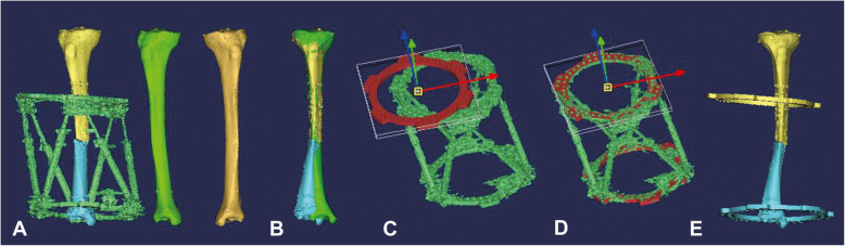
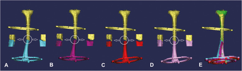
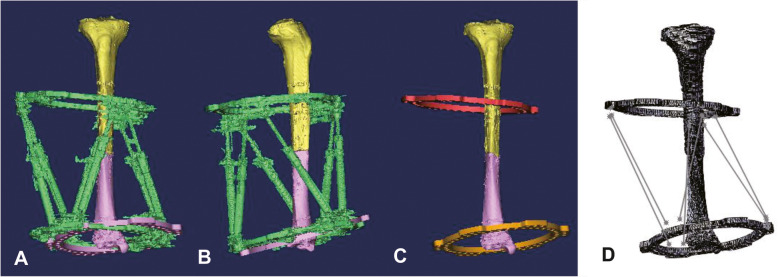
Methods: This retrospective study consists of 25 patients with high-energy tibial diaphyseal fractures treated by the HEF at our institution from January 2016 to June 2018, including 22 males and 3 females with a mean age of 42 years (range 14-63 years). Hexapod external fixator treatments were conducted to manage the multiplanar posttraumatic deformities with/without poor soft-tissue that were not suitable for internal fixation. Computer-assisted 3D reconstruction and trajectory planning of the reduction by Mimics were applied to perform virtual fracture reduction and deformity correction. The electronic prescription derived from the length changes of the six struts were calculated by SolidWorks. Fracture reduction was conducted by adjusting the lengths of the six struts according to the electronic prescription. Effectiveness was evaluated by the standard anteroposterior (AP) and lateral X-rays after reduction.
Results: All patients acquired excellent functional reduction and achieved bone union in our study. After correction, the mean translation (1.0 ± 1.1 mm) and angulation (0.8 ± 1.2°) on the coronal plane, mean translation (0.8 ± 1.0 mm) and angulation (0.3 ± 0.8°) on the sagittal plane were all less than those (6.1 ± 4.9 mm, 5.2 ± 3.2°, 4.2 ± 3.5 mm, 4.0 ± 2.5°) before correction (P < 0.05).
Conclusions: The computer-assisted three-dimensional reconstruction and hexapod external fixator-based method allows surgeons to conduct long bone fracture reduction and deformity correction without calculating the parameters needed by the traditional usage. This method is suggested to apply in those unusually complex cases with extensive soft tissue damage and where internal fixation is impossible or inadvisable.
Keywords: Computer‐assisted; Fracture reduction; Hexapod external fixation; Taylor spatial frame; Three‐dimensional reconstruction.
Conflict of interest statement
The authors declare that they have no conflict of interest.
Figures


Similar articles
-
Correction outcomes of the postoperative malalignment salvaged by the temporary application of the hexapod external fixator in tibial diaphyseal fractures treated by monolateral external fixation.Injury. 2021 Nov;52(11):3478-3482. doi: 10.1016/j.injury.2021.01.018. Epub 2021 Jan 14. Injury. 2021. PMID: 33487408
-
Application of elliptic registration and three-dimensional reconstruction in the postoperative measurement of Taylor spatial frame parameters.Injury. 2020 Dec;51(12):2975-2980. doi: 10.1016/j.injury.2020.10.077. Epub 2020 Oct 17. Injury. 2020. PMID: 33268078
-
Optimizing Outcomes in Distal Tibial Deformity Correction: The Role of Supramalleolar Osteotomy with Computer-assisted Hexapod External Fixator.Orthop Surg. 2024 Sep;16(9):2173-2180. doi: 10.1111/os.14201. Epub 2024 Aug 19. Orthop Surg. 2024. PMID: 39161056 Free PMC article.
-
[Corrective osteotomies around the knee joint using hexapods].Oper Orthop Traumatol. 2024 Apr;36(2):83-95. doi: 10.1007/s00064-023-00836-4. Epub 2023 Nov 10. Oper Orthop Traumatol. 2024. PMID: 37947855 Review. German.
-
Use of Hexapod External Fixation in Limb Lengthening in Patients with Disproportionate Short Stature: A Systematic Review of the Last 20 Years.J Clin Med. 2025 Feb 8;14(4):1091. doi: 10.3390/jcm14041091. J Clin Med. 2025. PMID: 40004622 Free PMC article. Review.
Cited by
-
The Clinical Application of Double Taylor Spatial Frame in Segmental Tibial Fracture.Orthop Surg. 2024 Jun;16(6):1344-1355. doi: 10.1111/os.14045. Epub 2024 Apr 25. Orthop Surg. 2024. PMID: 38664223 Free PMC article.
-
Finite element analysis and clinical evaluation of cross locking external fixator configuration for distal third tibia fracture.Sci Rep. 2025 Apr 17;15(1):13310. doi: 10.1038/s41598-025-97090-4. Sci Rep. 2025. PMID: 40247025 Free PMC article.
-
Definition of a measurement technique for hexapod circular smart fixators' perioperative assembly parameters and investigation of alignment and correlation with postoperative measurements: a retrospective cohort study.BMC Musculoskelet Disord. 2024 Nov 20;25(1):933. doi: 10.1186/s12891-024-08056-y. BMC Musculoskelet Disord. 2024. PMID: 39563261 Free PMC article.
References
-
- Green SA. Complications of external skeletal fixation. Clin Orthop Relat Res. 1983;180:109–16. - PubMed
MeSH terms
Grants and funding
LinkOut - more resources
Full Text Sources
Other Literature Sources
Medical
