

Cardiac involvement in COVID-19 patients: mid-term follow up by cardiovascular magnetic resonance
- PMID: 33627143
- PMCID: PMC7904320
- DOI: 10.1186/s12968-021-00710-x
Cardiac involvement in COVID-19 patients: mid-term follow up by cardiovascular magnetic resonance
Abstract
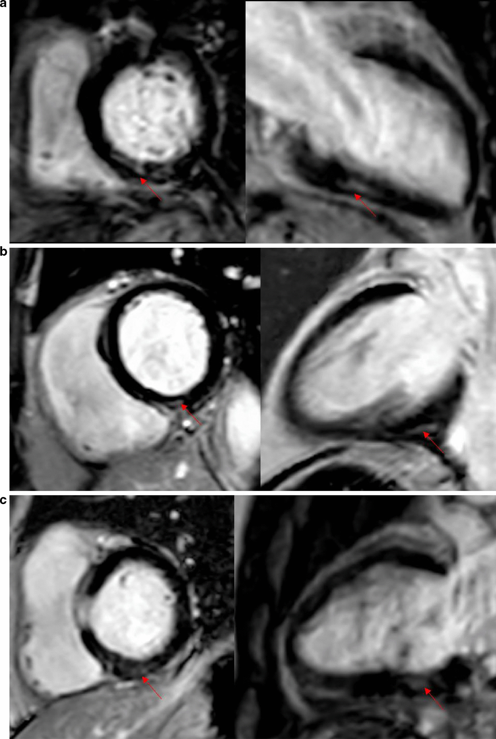
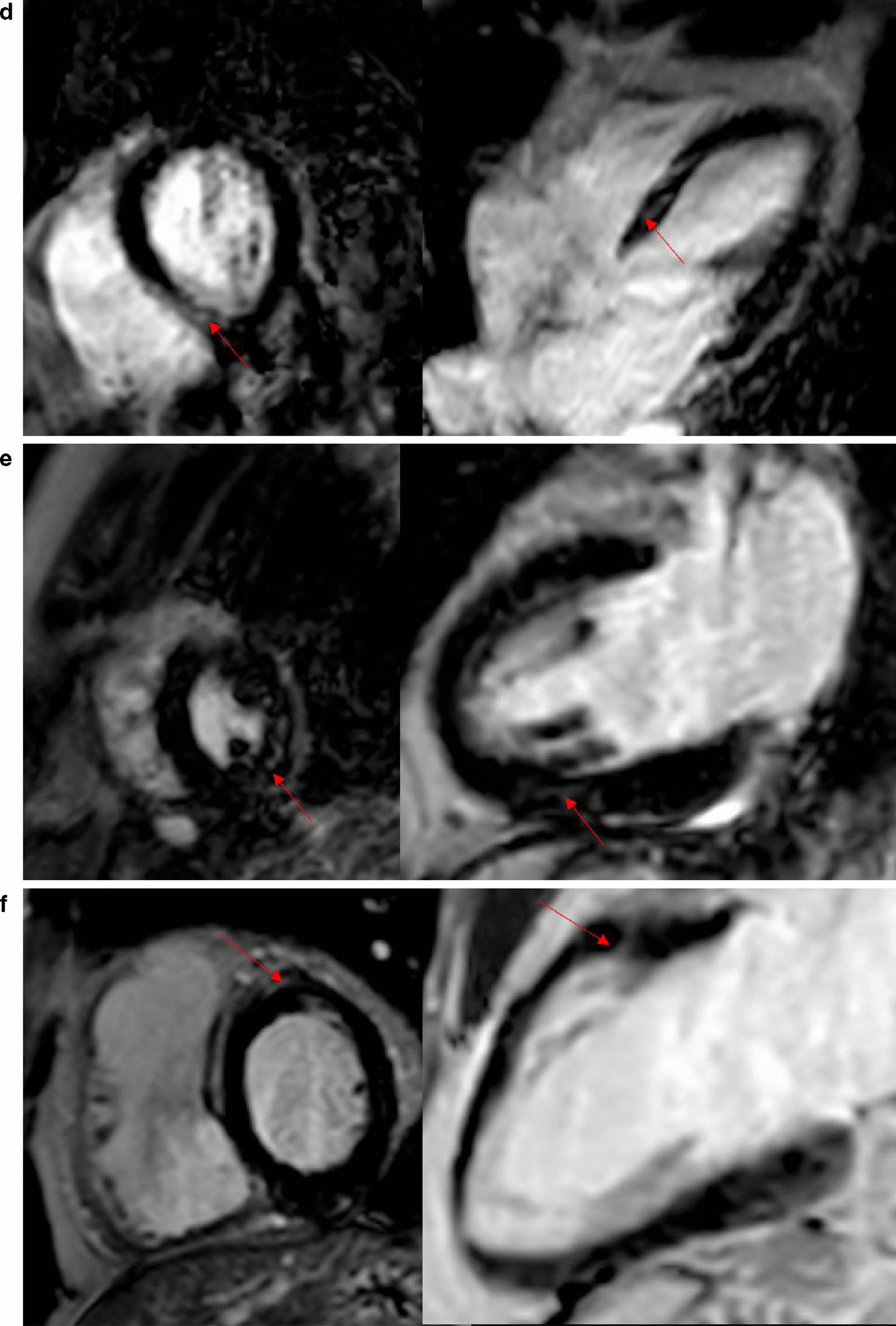
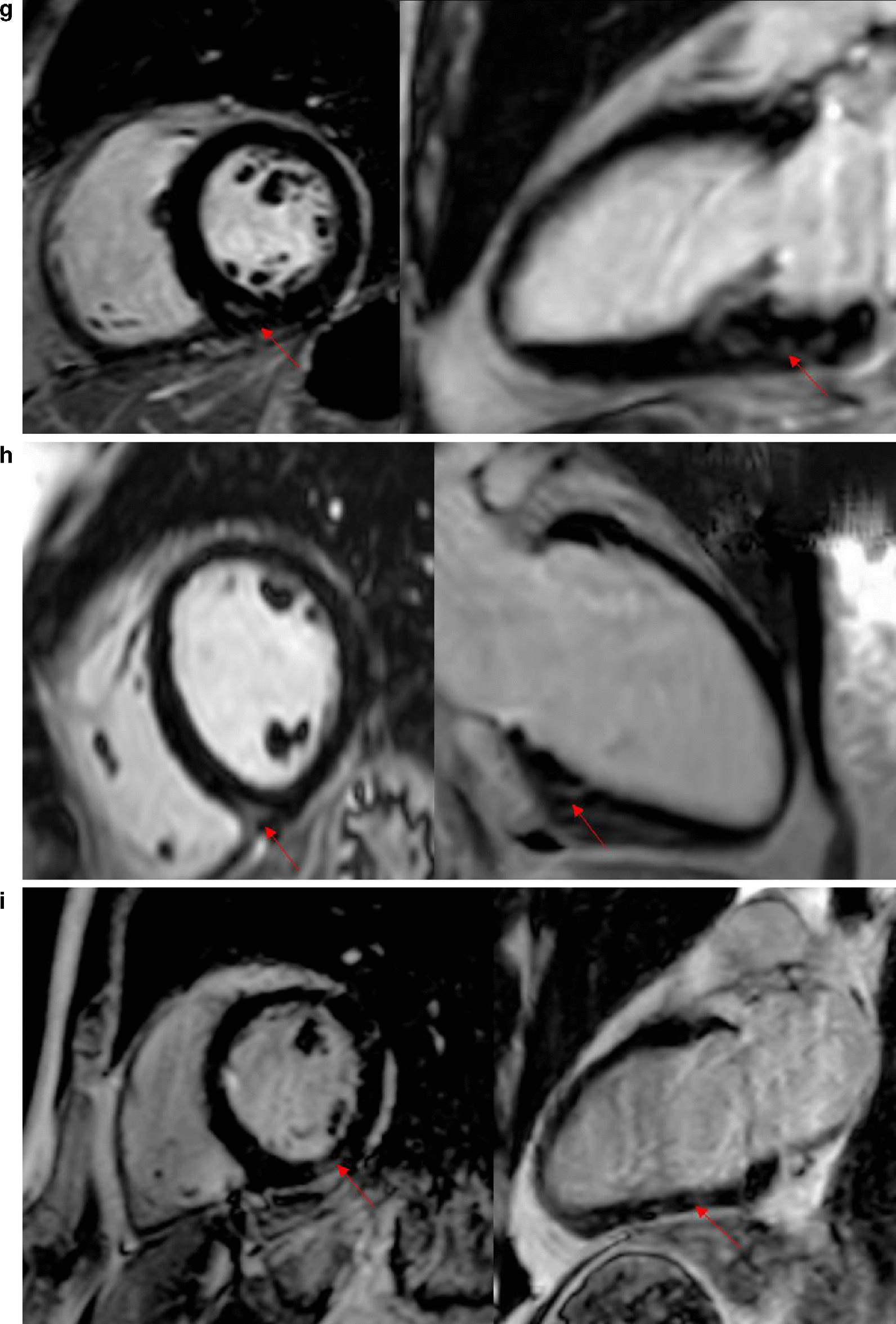
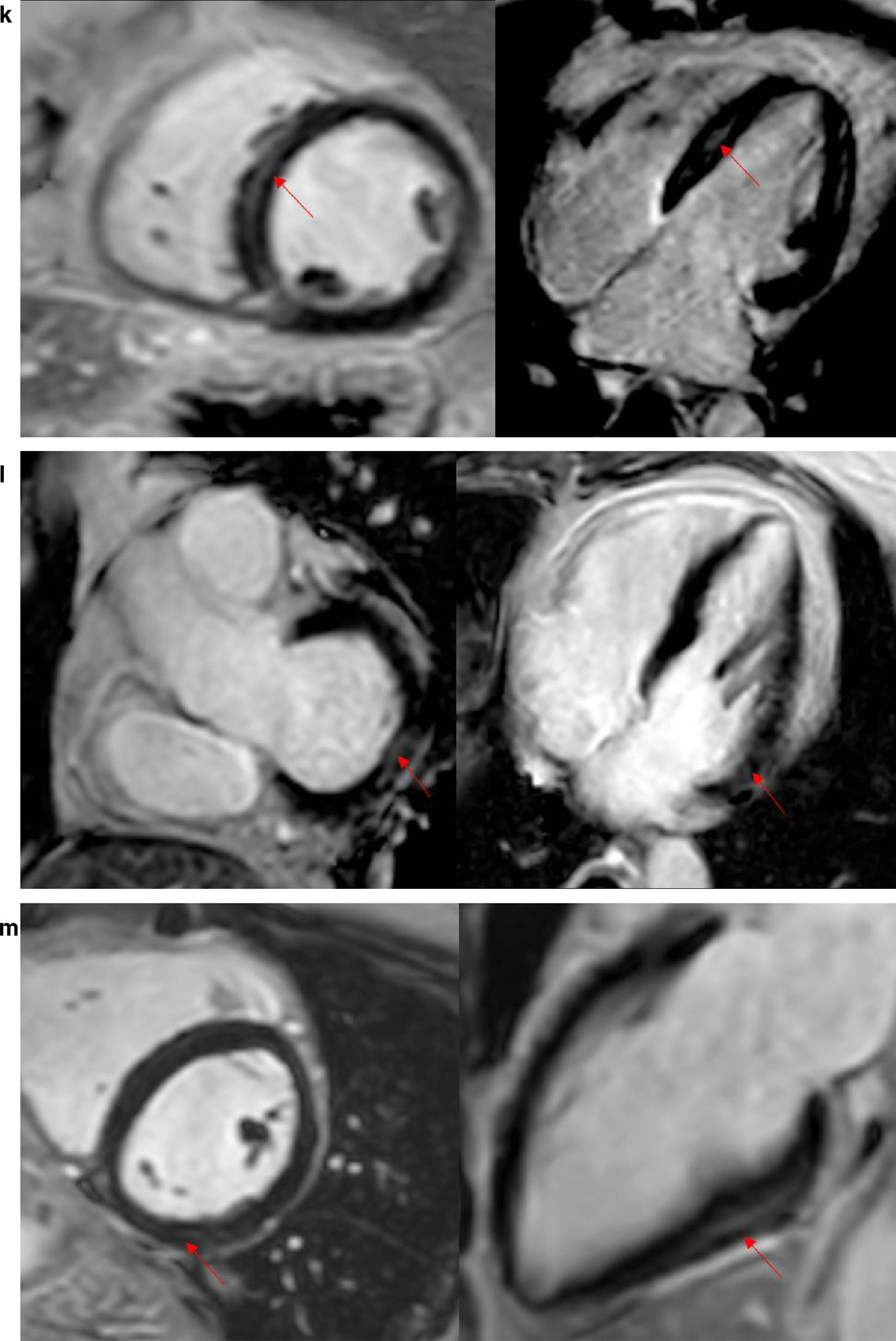
Background: Coronavirus disease 2019 (COVID-19) induces myocardial injury, either direct myocarditis or indirect injury due to systemic inflammatory response. Myocardial involvement has been proved to be one of the primary manifestations of COVID-19 infection, according to laboratory test, autopsy, and cardiovascular magnetic resonance (CMR). However, the middle-term outcome of cardiac involvement after the patients were discharged from the hospital is yet unknown. The present study aimed to evaluate mid-term cardiac sequelae in recovered COVID-19 patients by CMR METHODS: A total of 47 recovered COVID-19 patients were prospectively recruited and underwent CMR examination. The CMR protocol consisted of black blood fat-suppressed T2 weighted imaging, T2 star mapping, left ventricle (LV) cine imaging, pre- and post-contrast T1 mapping, and late gadolinium enhancement (LGE). LGE were assessed in mixed both recovered COVID-19 patients and healthy controls. The LV and right ventricle (RV) function and LV mass were assessed and compared with healthy controls.
Results: A total of 44 recovered COVID-19 patients and 31 healthy controls were studied. LGE was found in 13 (30%) of COVID-19 patients. All LGE lesions were located in the mid myocardium and/or sub-epicardium with a scattered distribution. Further analysis showed that LGE-positive patients had significantly decreased LV peak global circumferential strain (GCS), RV peak GCS, RV peak global longitudinal strain (GLS) as compared to non-LGE patients (p < 0.05), while no difference was found between the non-LGE patients and healthy controls.
Conclusion: Myocardium injury existed in 30% of COVID-19 patients. These patients have depressed LV GCS and peak RV strains at the 3-month follow-up. CMR can monitor the COVID-19-induced myocarditis progression, and CMR strain analysis is a sensitive tool to evaluate the recovery of LV and RV dysfunction.
Keywords: COVID-19; Cardiac dysfunction; Cardiac involvement; Cardiac magnetic resonance imaging.
Conflict of interest statement
The authors declare that they have no competing interests.
Figures

References
-
- WHO coronavirus disease (COVID-19) dashboard. Geneva: World Health Organization, 2020. https://covid19.who.int/. In. 2020/7/4 edn.
Publication types
MeSH terms
Grants and funding
- U1908211/National Natural Science Foundation of China
- 2016YFC1300300/National Key Research and Development Program of China
- PXM2020_026272_000013/Capital Medical Development Research Foundation of China
- 2020-TG-002/Beijing Municipal Health Commission, technology achievements and appropriate technology promotion project
- BJYAYY-2020YC-03/You'an Medical Development Project of COVID-19 Emergency Prevention and Control Public
LinkOut - more resources
Full Text Sources
Other Literature Sources
Medical
Miscellaneous
