. 2021 May;70(5):1170-1184.
doi: 10.2337/db20-1008. Epub 2021 Feb 24.

Dedicator of Cytokinesis 5 Regulates Keratinocyte Function and Promotes Diabetic Wound Healing

Affiliations

Dedicator of Cytokinesis 5 Regulates Keratinocyte Function and Promotes Diabetic Wound Healing

Hua Qu et al. Diabetes. 2021 May.

Abstract

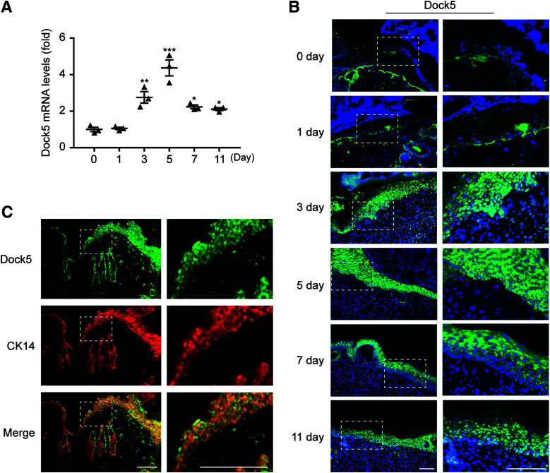
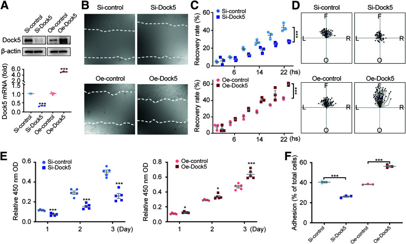
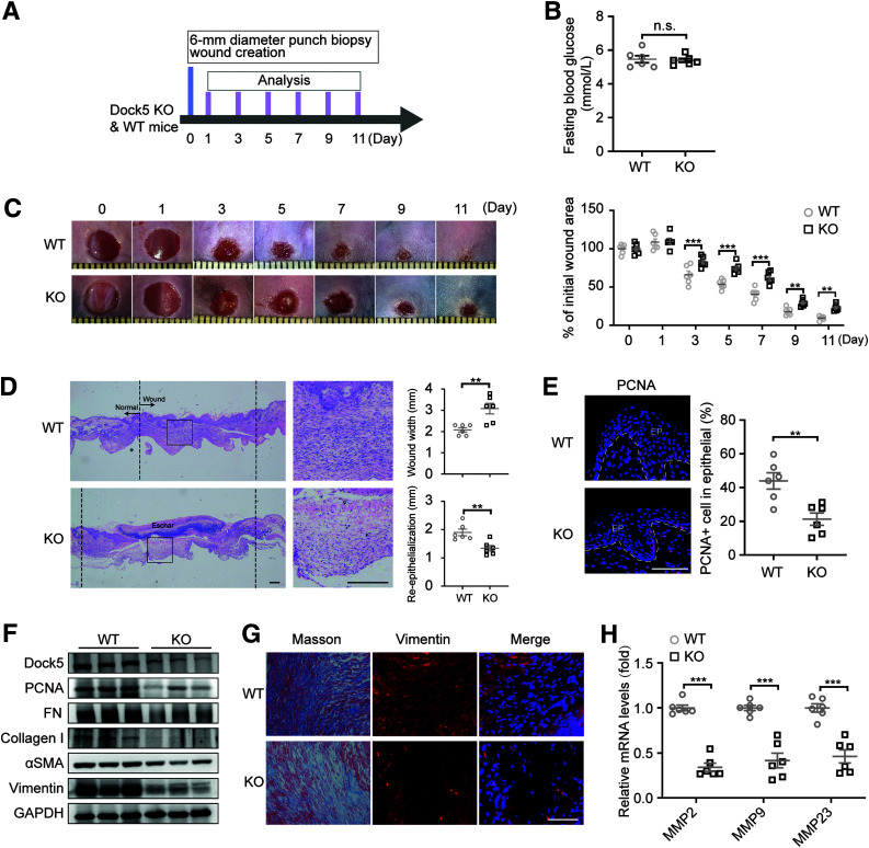
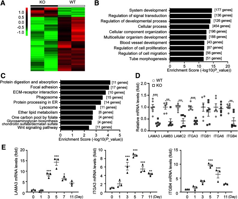
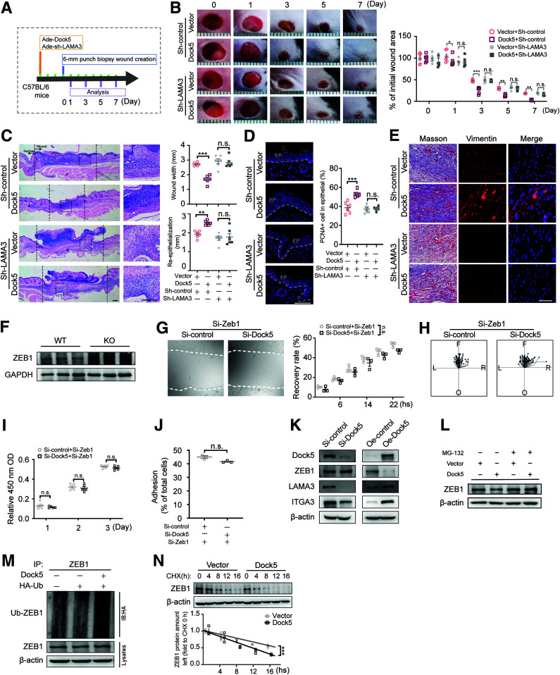
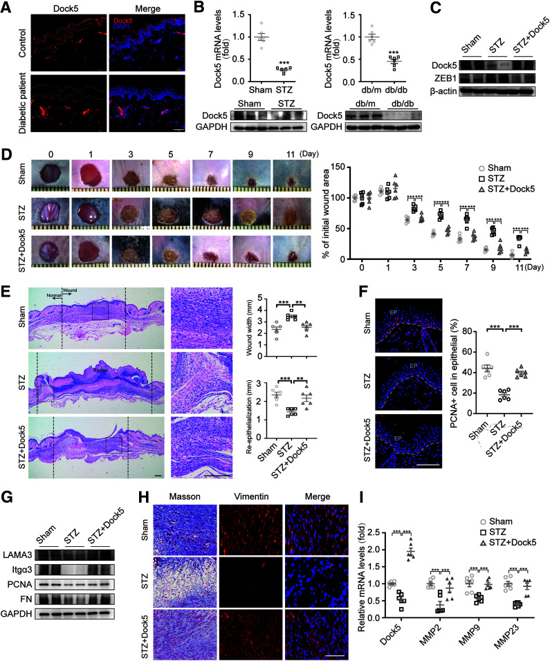
Cutaneous wound healing is a fundamental biologic and coordinated process, and failure to maintain this process contributes to the dysfunction of tissue homeostasis, increasing the global burden of diabetic foot ulcerations. However, the factors that mediate this process are not fully understood. Here, we identify the pivotal role of dedicator of cytokinesis 5 (Dock5) in keratinocyte functions contributing to the process of skin wound healing. Specifically, Dock5 is highly upregulated during the proliferative phase of wound repair and is predominantly expressed in epidermal keratinocytes. It regulates keratinocyte adhesion, migration, and proliferation and influences the functions of extracellular matrix (ECM) deposition by facilitating the ubiquitination of transcription factor ZEB1 to activate laminin-332/integrin signaling. Genetic ablation of Dock5 in mice leads to attenuated reepithelialization and granulation tissue formation, and Dock5 overexpression-improved skin repair can be abrogated by LAMA3 knockdown. Importantly, Dock5 expression in the skin edge is reduced in patients and animal models of diabetes, further suggesting a direct correlation between its abundance and healing capability. The rescue of Dock5 expression in diabetic mice causes a significant improvement in reepithelialization, collagen deposition, ECM production, and granulation. Our study provides a potential therapeutic target for wound healing impairment during diabetes.


Figures

Figure 1
Figure 1
Expression pattern of Dock5 in wounds. Full-thickness wounds were made on the dorsum of C57BL/6 mice. A: mRNA expression of Dock5 in wound biopsies at the indicated time points after injury was analyzed by quantitative real-time PCR. B: In situ hybridization was performed using a Dock5-specific probe. Green indicates Dock5 expression, and blue indicates DAPI. C: Wound sections from C57BL/6 mice were stained for Dock5 (green) and cytokeratin 14 (CK14) (red, keratinocyte marker). Dotted lines in B and C indicate magnified area. Scale bars, 100 μm for B and C. n = 3 mice per group for A–C. Data are presented as means ± SEMs. *P < 0.05, **P < 0.01, ***P < 0.001.
Figure 2
Figure 2
Dock5 regulates keratinocyte migration, proliferation, and adhesion. Keratinocytes were transfected with Dock5-specific siRNA (Si-Dock5) or overexpression plasmid (Oe-Dock5). A: The transfection efficiency of overexpression and knockdown is presented. B: Live cell images were acquired 16 h after scratching in a wound-healing assay via a live cell imaging system. C: Wound recovery rate was quantified. D: Cell migration trajectory was tracked for 2 h at 8–10 h after scratching. The center indicates the start point, and magenta dots indicate end points. Each line shows the trajectory of a randomly chosen cell (left [L], right [R], forward [F], or opposite [O] of the direction of migration). E: Cell proliferation was assessed by Cell Counting Kit 8 assay. F: Cell adhesion was quantified using the CyQUANT Assay Kit. n = 3 for A–C and F, n = 65–75 for D, and n = 5 mice per group for E. Data are presented as means ± SEMs. *P < 0.05, ***P < 0.001. OD, optical density.
Figure 3
Figure 3
Dock5 KO impedes wound healing. Full-thickness wounds were made on the dorsum of Dock5 KO and WT mice. A: Experiment setup. B: Fasting blood glucose was assessed. C: Representative wound images (left) are shown at the indicated times and were quantified as the percentages of the initial wound area (right). D: Representative hematoxylin-eosin–stained sections are shown on day 7 after injury, and wound width and reepithelialization were quantified in WT and KO mice. E: The representative images and quantification of PCNA+ cell numbers in epithelial cells are shown. Epithelium (EP) is tagged with dotted lines. F: Expression of the indicated proteins in wound tissue from WT and KO mice by Western blot analysis. G: Masson trichrome staining and vimentin immunostaining of wound sections from WT and KO mice. H: mRNA expression of genes related to ECM deposition was analyzed by quantitative real-time PCR. Each grid scale represents 1 mm for C. Scale bars, 200 μm for D and 50 μm for E and G. n = 6 mice per group for A–H. Data are presented as means ± SEMs. **P < 0.01, ***P < 0.001. FN, fibronectin; n.s., not significant.
Figure 4
Figure 4
Dock5 regulates laminin-332/integrin signaling during wound healing. A: Hierarchic clustering of all significantly differentially expressed genes in wound tissue at day 5 after injury. B: Gene ontology analysis of all significantly altered genes was performed, and the top 10 items are shown. C: KEGG pathway analysis was conducted on all significantly downregulated genes, and the top 10 items are shown. D: Gene expression of all three chains of laminin-332 and its two integrin receptors in wound tissue from WT and KO mice was assessed by quantitative real-time PCR (qRT-PCR). E: mRNA expression of indicated genes in wound biopsies at the indicated time points after injury was analyzed by qRT-PCR. n = 6 mice per group for D, and n = 3 for E. Data are presented as means ± SEMs. *P < 0.05, **P < 0.01, ***P < 0.001. ER, endoplasmic reticulum.
Figure 5
Figure 5
Dock5 accelerates wound healing by LAMA3 activation through promoting ZEB1 ubiquitination. A: Schematic representation of the experiment setup used for evaluating whether Dock5-regulated wound healing occurs through LAMA3. Dock5-expressing and LAMA3 knockdown adenoviruses (Ade) were intradermally injected around the wound areas 3 days before the full-thickness wounds were made in C57BL/6 mice. B: Representative wound images (left) are shown at the indicated times and quantified as the percentages of the initial wound area (right). C: Representative hematoxylin-eosin–stained sections (left) are shown on day 7 after injury, and wound width and reepithelialization (right) were quantified. D: The representative images and quantification of PCNA+ cell numbers in epithelial cells are shown. Epithelium (EP) is tagged with dotted lines. E: Masson trichrome staining and vimentin immunostaining of wound sections from indicated mice. F: Expression of the ZEB1 proteins in wound tissue from WT and KO mice by Western blot analysis. Keratinocytes were transfected with Zeb1-specific siRNA with or without Dock5-specific siRNA. G: Live cell images (left) were acquired 16 h after scratching in a wound-healing assay via a live cell imaging system, and the wound recovery rate was quantified (right). H: Cell migration trajectories were tracked for 2 h at 8–10 h after scratching. The center indicates the start point, and magenta dots indicate end points. Each line shows the trajectory of a randomly chosen cell (left [L], right [R], forward [F], or opposite [O] of the direction of migration). I: Cell proliferation was assessed by Cell Counting Kit 8 assay. J: Cell adhesion was quantified using the CyQUANT Assay Kit. K: Keratinocytes were transfected with Dock5-specific siRNA or Dock5 overexpression plasmid, and indicated proteins were detected. L–N: Keratinocytes were transfected by Dock5 plasmid, and ZEB1 protein was examined 4 h after proteasomal inhibitor MG-132 (10 μmol/L) addition (L). Hemagglutinin (HA)-tagged ubiquitin (Ub) was cotransfected with or without Dock5 plasmid, and ubiquitination was detected (M). The half-life of ZEB1 protein was determined by pulse-chase assay with protein synthesis inhibitor cycloheximide (CHX) (25 μmol/L) administration (N). Each grid scale represents 1 mm for B. Scale bars, 200 μm for C, and 50 μm for D and E. n = 3 mice per group for G and J–N, n = 5 for I, n = 69–75 for H, n = 6 for A–F. Data are presented as means ± SEMs. *P < 0.05, **P < 0.01, ***P < 0.001. IB, immunoblotting; IP, immunoprecipitation; n.s., not significant; OD, optical density.
Figure 6
Figure 6
Rescue of Dock5 accelerates wound healing in diabetic mice. A: Skin tissue surrounding the wounds of patients with diabetes was sampled and immunostained for Dock5. Epithelium (EP) is tagged with dotted lines. B: mRNA and protein levels of Dock5 were analyzed in wound tissue of STZ and db/db mice. Full-thickness wounds were made on the dorsum of control (Sham) and STZ-injected mice, and Dock5-expressing adenovirus was then intradermally injected into each wound of the STZ group mice 3 days before the wounds were created. C: The protein expression of Dock5 and ZEB1 was assessed in isolated keratinocytes from wound tissues. D: Representative wound images (left) are shown at the indicated times, and healing was quantified as the percentage of the initial wound area (right). E: Representative hematoxylin-eosin–stained sections (left) are shown on day 7 after injury, and the wound width and reepithelialization (right) were quantified. F: The representative images and quantification of PCNA+ cell numbers in epithelial cells are shown for each group. G: Expression of the indicated proteins in wound tissue from mice by Western blot analysis. H: Masson trichrome staining and vimentin immunostaining of wound sections are shown for the indicated groups. I: mRNA expression of genes related to ECM deposition was analyzed by quantitative real-time PCR. Each grid scale represents 1 mm for D. Scale bars, 100 μm for A, 200 μm for E, and 50 μm for F and H. n = 6 patients or mice per group for A–I. Data are presented as means ± SEMs. **P < 0.01, ***P < 0.001.

Similar articles

Cited by

References

    1. Eming SA, Martin P, Tomic-Canic M.. Wound repair and regeneration: mechanisms, signaling, and translation. Sci Transl Med 2014;6:265sr6PubMed - PMC - PubMed
    1. Raja, Sivamani K, Garcia MS, Isseroff RR.. Wound re-epithelialization: modulating keratinocyte migration in wound healing. Front Biosci 2007;12:2849–2868 - PubMed
    1. Simpson CL, Patel DM, Green KJ.. Deconstructing the skin: cytoarchitectural determinants of epidermal morphogenesis. Nat Rev Mol Cell Biol 2011;12:565–580PubMed - PMC - PubMed
    1. Watt FM. Mammalian skin cell biology: at the interface between laboratory and clinic. Science 2014;346:937–940PubMed - PubMed
    1. Longmate WM, Dipersio CM.. Integrin regulation of epidermal functions in wounds. Adv Wound Care (New Rochelle) 2014;3:229–246PubMed - PMC - PubMed

Publication types

MeSH terms