. 2021 Apr 12;95(9):e02359-20.
doi: 10.1128/JVI.02359-20. Print 2021 Apr 12.

Effect of an Adenovirus-Vectored Universal Influenza Virus Vaccine on Pulmonary Pathophysiology in a Mouse Model

Affiliations

Effect of an Adenovirus-Vectored Universal Influenza Virus Vaccine on Pulmonary Pathophysiology in a Mouse Model

Santosh Dhakal et al. J Virol. .

Abstract

Current influenza vaccines, live attenuated or inactivated, do not protect against antigenically novel influenza A viruses (IAVs) of pandemic potential, which has driven interest in the development of universal influenza vaccines. Universal influenza vaccine candidates targeting highly conserved antigens of IAV nucleoprotein (NP) are promising as vaccines that induce T cell immunity, but concerns have been raised about the safety of inducing robust CD8 T cell responses in the lungs. Using a mouse model, we systematically evaluated effects of recombinant adenovirus vectors (rAd) expressing IAV NP (A/NP-rAd) or influenza B virus (IBV) NP (B/NP-rAd) on pulmonary inflammation and function after vaccination and following live IAV challenge. After A/NP-rAd or B/NP-rAd vaccination, female mice exhibited robust systemic and pulmonary vaccine-specific B cell and T cell responses and experienced no morbidity (e.g., body mass loss). Both in vivo pulmonary function testing and lung histopathology scoring revealed minimal adverse effects of intranasal rAd vaccination compared with unvaccinated mice. After IAV challenge, A/NP-rAd-vaccinated mice experienced significantly less morbidity, had lower pulmonary virus titers, and developed less pulmonary inflammation than unvaccinated or B/NP-rAd-vaccinated mice. Based on analysis of pulmonary physiology using detailed testing not previously applied to the question of T cell damage, mice protected by vaccination also had better lung function than controls. Results provide evidence that, in this model, adenoviral universal influenza vaccine does not damage pulmonary tissue. In addition, adaptive immunity, in particular, T cell immunity in the lungs, does not cause damage when restimulated but instead mitigates pulmonary damage following IAV infection.IMPORTANCE Respiratory viruses can emerge and spread rapidly before vaccines are available. It would be a tremendous advance to use vaccines that protect against whole categories of viruses, such as universal influenza vaccines, without the need to predict which virus will emerge. The nucleoprotein (NP) of influenza virus provides a target conserved among strains and is a dominant T cell target. In animals, vaccination to NP generates powerful T cell immunity and long-lasting protection against diverse influenza strains. Concerns have been raised, but not evaluated experimentally, that potent local T cell responses might damage the lungs. We analyzed lung function in detail in the setting of such a vaccination. Despite CD8 T cell responses in the lungs, lungs were not damaged and functioned normally after vaccination alone and were protected upon subsequent infection. This precedent provides important support for vaccines based on T cell-mediated protection, currently being considered for both influenza and SARS-CoV-2 vaccines.

Keywords: T cell response; adenovirus; antibody; influenza A virus; influenza B virus; oxygen exchange; pulmonary function; recombinant adenovirus; universal influenza vaccine; vaccine safety.


Figures

FIG 1
FIG 1
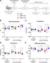
Adenoviral universal influenza vaccine does not alter pulmonary function and lung integrity after vaccination. (A) Adult (8- to 10-week-old) female mice were vaccinated once with A/NP-rAd, B/NP-rAd, or PBS by the intranasal route. Six days after vaccination, a subset of mice was utilized to measure pulmonary functions and euthanized for performing histopathology analysis of lung tissues. (B to E) Total lung capacity (B), pulmonary compliance (C), pulmonary resistance (D), and lung diffusion capacity (E) were measured for determining lung function. (F and G) Representative images of H&E-stained sections from each group taken at ×10 magnification (F) and cumulative inflammation scores in the lungs (G) are shown. Data represent mean ± standard error of the mean (SEM) from 7 to 8 mice/group, and significant differences (*, P < 0.05) between groups are denoted by asterisks based on one-way ANOVAs.
FIG 2
FIG 2
Single-dose intranasal adenoviral universal influenza vaccine induces antibody and T cell immunity. (A) Adult (8- to 10-week-old) female mice were vaccinated once with A/NP-rAd, B/NP-rAd, or PBS by the intranasal route. Twenty-eight days after vaccination, serum was collected to analyze antibody responses, while a subset of mice (n = 3/group) was euthanized at 34 days after vaccination to analyze T cell immunity in the lungs and IgA antibody responses in BAL fluid. (B to E) IgG antibody responses against A/NP (B) and B/NP (C) and IgA antibody responses against A/NP (D) and B/NP (E) were determined by ELISA. (F) T cell responses were measured by ELISPOT detecting IFN-γ-secreting cells in response to stimulation by indicated peptides. Data represent the average number of IFN-γ-positive lung cells per 106 cells from 3 mice per immunization group, with each sample run in triplicate. Bars show mean ± SEM for three animals. Converting to total responding lung cells per animal, results correspond to, for example, a CD8 response to the NP366 epitope in the lungs of A/NP-immunized mice of approximately 25,000 cells/mouse.
FIG 3
FIG 3
Single-dose intranasal adenoviral universal influenza vaccine protects mice from influenza virus infection. (A) Adult (8- to 10-week-old) female mice were vaccinated once with A/NP-rAd, B/NP-rAd, or PBS by the intranasal route. At 35 days after vaccination, mice were challenged with mouse-adapted 2009 H1N1 virus (102 TCID50). A subset of mice (n = 8/group) was euthanized at 7 days postchallenge to determine virus titer in the lungs, and another subset (n = 8 to 9/group) was followed for 14 days for morbidity measures. (B to D) The infectious virus titer in the lungs at 7 days postchallenge (B), percentage change in the body mass (C), and rectal temperature (D) until 14 days postchallenge are shown. One of the 9 mice in PBS group reached the humane endpoint of 30% body mass loss on day 11 postchallenge, while all other mice recovered. Data represent mean ± standard error of the mean from 8 to 9 mice/group. In panel B, significant differences (*, P < 0.05) between groups are denoted by asterisks as determined by one-way ANOVA. Likewise, * and # in panels C and D represent significant differences between A/NP-rAd versus PBS and A/NP-rAd versus B/NP-rAd, respectively, based on repeated-measures ANOVA (mixed model).
FIG 4
FIG 4
Adenoviral universal influenza vaccine maintains pulmonary functions after influenza virus challenge. (A) Adult (8- to 10-week-old) female mice were vaccinated once with A/NP-rAd, B/NP-rAd, or PBS by the intranasal route. At 35 days after vaccination, mice were challenged with mouse-adapted 2009 H1N1 virus (102 TCID50). Pulmonary function tests were performed at 7 and 14 days postchallenge. (B to E) Total lung capacity (B), pulmonary compliance (C), pulmonary resistance (D), and lung diffusion capacity (E) were measured to determine lung functions. Data represent mean ± SEM from 6 to 8 mice/group, and significant differences (*, P < 0.05) between groups are denoted by asterisks based on two-way ANOVAs.
FIG 5
FIG 5
Adenoviral universal influenza vaccine does not exacerbate pulmonary inflammation after subsequent infection with influenza virus. Adult (8- to 10-week-old) female mice were vaccinated once with A/NP-rAd, B/NP-rAd, or PBS by the intranasal route. At 35 days after vaccination, mice were challenged with mouse-adapted 2009 H1N1 virus (102 TCID50). A subset of mice was euthanized at 7 and 14 days postchallenge, and lungs were collected, fixed, and H&E stained. (A) Representative images of H&E-stained sections taken at ×4 magnification are shown and can be compared to the prechallenge histopathology images in Fig. 1F. (B to E) Cumulative pulmonary inflammation (B) was determined on a scale of 0 to 3 each for perivasculitis (C), peribronchiolitis (D), and alveolitis (E). Data represent mean ± SEM from 8 mice/group, and significant differences between groups are denoted by asterisks (*, P < 0.05) based on two-way ANOVAs.

References

    1. Erbelding EJ, Post D, Stemmy E, Roberts PC, Augustine AD, Ferguson S, Paules CI, Graham BS, Fauci AS. 2018. A universal influenza vaccine: the strategic plan for the National Institute of Allergy and Infectious Diseases. J Infect Dis 218:347–354. 10.1093/infdis/jiy103. - DOI - PMC - PubMed
    1. Epstein SL. 2018. Universal influenza vaccines: progress in achieving broad cross-protection in vivo. Am J Epidemiol 187:2603–2614. 10.1093/aje/kwy145. - DOI - PMC - PubMed
    1. Okuno Y, Matsumoto K, Isegawa Y, Ueda S. 1994. Protection against the mouse-adapted A/FM/1/47 strain of influenza A virus in mice by a monoclonal antibody with cross-neutralizing activity among H1 and H2 strains. J Virol 68:517–520. 10.1128/JVI.68.1.517-520.1994. - DOI - PMC - PubMed
    1. Ekiert DC, Bhabha G, Elsliger MA, Friesen RH, Jongeneelen M, Throsby M, Goudsmit J, Wilson IA. 2009. Antibody recognition of a highly conserved influenza virus epitope. Science 324:246–251. 10.1126/science.1171491. - DOI - PMC - PubMed
    1. Kallewaard NL, Corti D, Collins PJ, Neu U, McAuliffe JM, Benjamin E, Wachter-Rosati L, Palmer-Hill FJ, Yuan AQ, Walker PA, Vorlaender MK, Bianchi S, Guarino B, De Marco A, Vanzetta F, Agatic G, Foglierini M, Pinna D, Fernandez-Rodriguez B, Fruehwirth A, Silacci C, Ogrodowicz RW, Martin SR, Sallusto F, Suzich JA, Lanzavecchia A, Zhu Q, Gamblin SJ, Skehel JJ. 2016. Structure and function analysis of an antibody recognizing all influenza A subtypes. Cell 166:596–608. 10.1016/j.cell.2016.05.073. - DOI - PMC - PubMed

Publication types

MeSH terms

Substances