. 2021 Mar 2;118(9):e2020606118.
doi: 10.1073/pnas.2020606118.

Generating tumor-selective conditionally active biologic anti-CTLA4 antibodies via protein-associated chemical switches

Affiliations

Generating tumor-selective conditionally active biologic anti-CTLA4 antibodies via protein-associated chemical switches

Hwai Wen Chang et al. Proc Natl Acad Sci U S A. .

Abstract

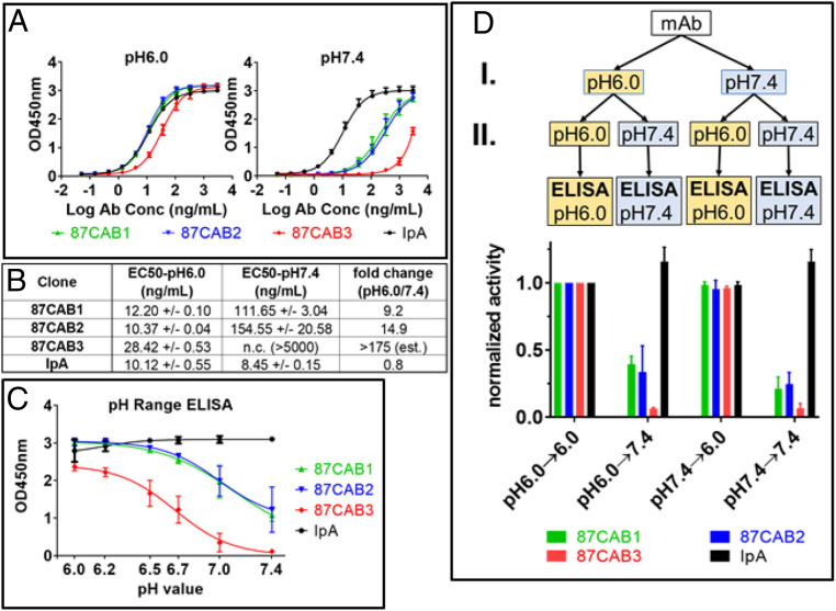
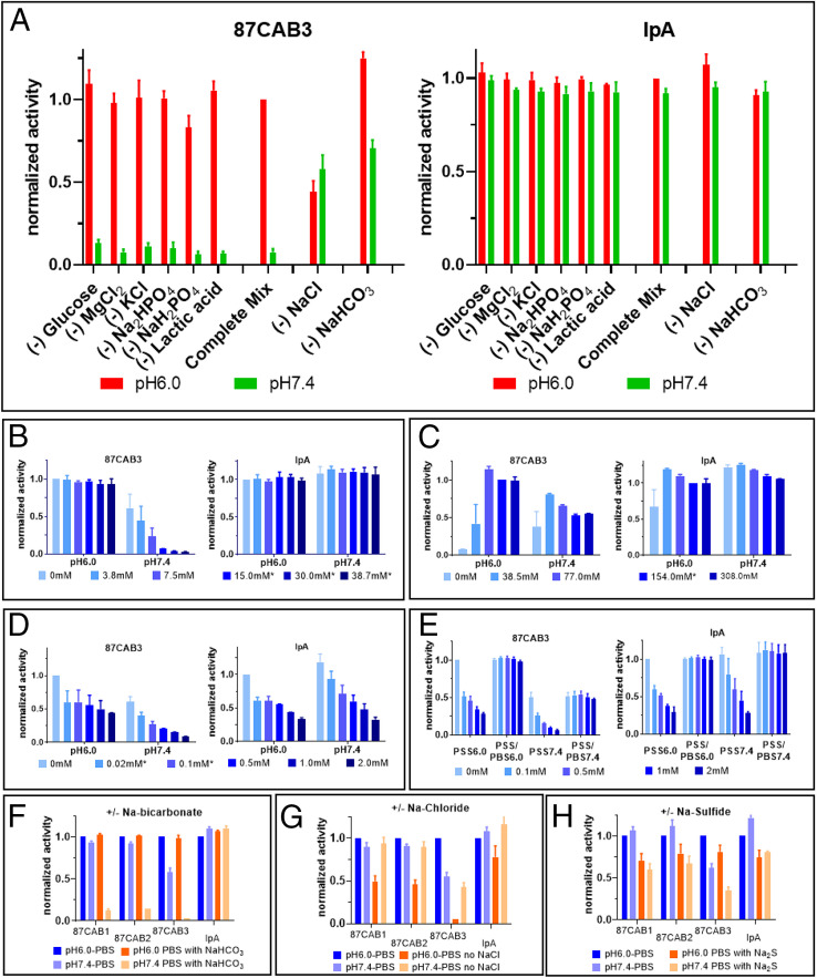
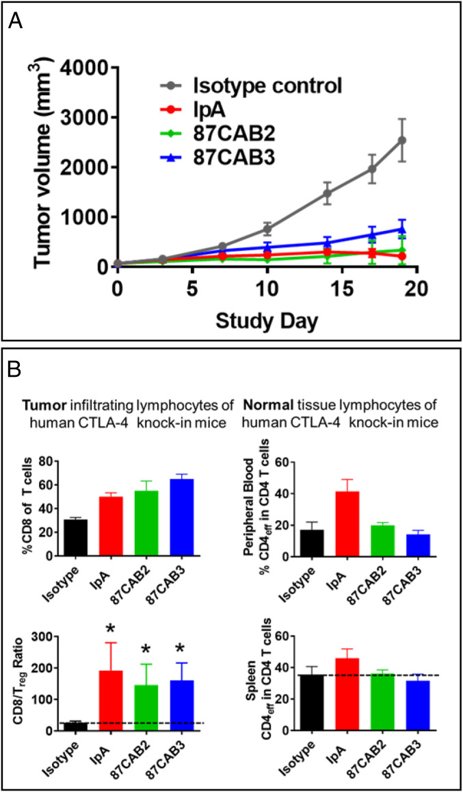
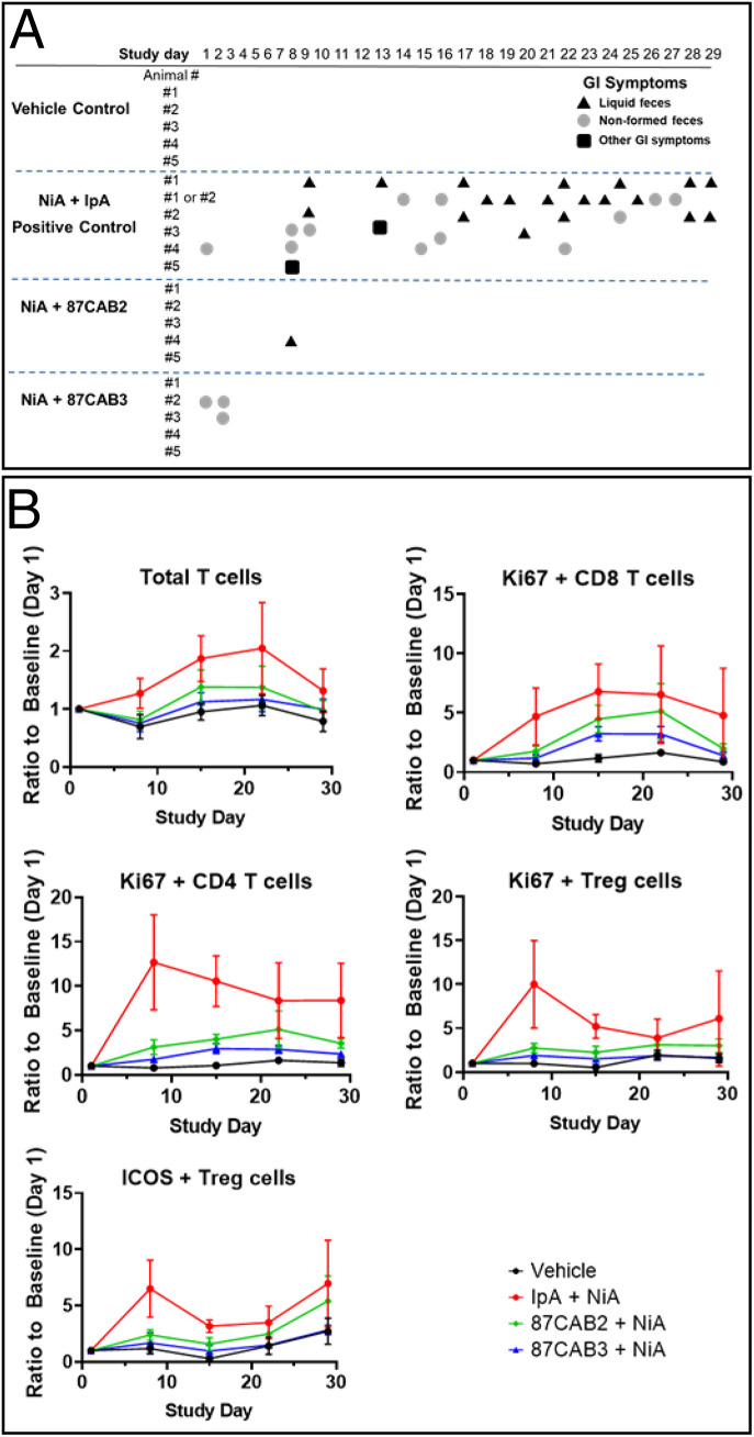
Anticytotoxic T lymphocyte-associated protein 4 (CTLA4) antibodies have shown potent antitumor activity, but systemic immune activation leads to severe immune-related adverse events, limiting clinical usage. We developed novel, conditionally active biologic (CAB) anti-CTLA4 antibodies that are active only in the acidic tumor microenvironment. In healthy tissue, this binding is reversibly inhibited by a novel mechanism using physiological chemicals as protein-associated chemical switches (PaCS). No enzymes or potentially immunogenic covalent modifications to the antibody are required for activation in the tumor. The novel anti-CTLA4 antibodies show similar efficacy in animal models compared to an analog of a marketed anti-CTLA4 biologic, but have markedly reduced toxicity in nonhuman primates (in combination with an anti-PD1 checkpoint inhibitor), indicating a widened therapeutic index (TI). The PaCS encompass mechanisms that are applicable to a wide array of antibody formats (e.g., ADC, bispecifics) and antigens. Examples shown here include antibodies to EpCAM, Her2, Nectin4, CD73, and CD3. Existing antibodies can be engineered readily to be made sensitive to PaCS, and the inhibitory activity can be optimized for each antigen's varying expression level and tissue distribution. PaCS can modulate diverse physiological molecular interactions and are applicable to various pathologic conditions, enabling differential CAB antibody activities in normal versus disease microenvironments.

Keywords: CTLA4; conditionally active biologics; immunooncology; monoclonal antibodies; protein-associated chemical switches.


Conflict of interest statement

Competing interest statement: All authors are shareholders of BioAtla, Inc., which owns the intellectual property rights to CABs- and PaCS-related technologies. H.W.C., G.F., and J.M.S. are inventors on relevant patents. (L.S. is not an inventor on relevant patents.) L.S. and J.M.S. serve as Directors of BioAtla.

Figures

Fig. 1.
Fig. 1.
The pH selectivity of anti-CTLA4 variants. CAB anti-CTLA4 antibodies were identified by screening a library of antibody variants for binding to recombinant human CTLA4 in acidic pH, but not in neutral pH, using the PaCS process. (A) Binding activities of CAB anti-CTLA4 variants to human CTLA4 at pH6.0 (Left) and at pH7.4 (Right) were determined using pH affinity ELISA; y axis, optical density (OD) 450 nm; x axis, antibody concentration (log nanograms per milliliter); the starting concentration is 3 µg/mL. The mean OD values from two independent experiments with duplicate reactions are shown. (B) EC50 values of CAB anti-CTLA4 variants binding to human CTLA4 at pH6.0 and at pH7.4 were calculated using the nonlinear fit (variable slope, four parameters) model built into GraphPad Prism software version 7.03. The binding curve for clones 87CAB3 did not reach saturation at pH 7.4, and therefore the EC50 value could not be calculated. (C) Binding activities of CAB anti-CTLA4 variants to human CTLA4 at different pH values were determined by pH range ELISA; y axis, OD 450 nm; x axis, pH of the ELISA mix and wash buffers. The mean OD values from two independent experiments with two replicates for each pH test are shown. Inflection point of the pH curves: Mean OD values (from two independent experiments) at the different pH were plotted against the pH of the buffer using GraphPad Prism software version 7.03. Curve fitting was done using the four-parameter model built into the software. The inflection point of the pH curve (pH with 50% binding activity) equals IC50 of the fitting equation. Binding activity at pH6.0 was set to 100%; 87CAB1 pH inflection point, pH6.92; 87CAB2 inflection point, pH6.95; 87CAB3 pH inflection point, pH6.66. (D) The pH-dependent binding of CAB anti-CTLA4 variants is reversible. The CAB anti-CTLA4 variants (clones 87CAB1, yellow; 87CAB2, purple; and 87CAB3, red) were tested in four different conditions: Antibodies were first diluted from stock solutions (in phosphate-buffered saline [PBS] pH7.4) into pH6.0 or pH7.4 ELISA mix at 250 ng/mL and incubated at room temperature for 30 min (step I in the flowchart). The mAbs were then further diluted to 25 ng/mL in pH6.0 or pH7.4 ELISA mix (step II). Binding to human CTLA4 immobilized in the wells was tested by ELISA. All ELISA steps (blocking, incubation, and washing) were done in either pH6.0 or pH7.4 ELISA mix as indicated in the flowchart. Data from two independent experiments with two replicates were normalized to test condition I (pH6.0→6.0). IpA was used as non-CAB reference in all experiments (black).
Fig. 2.
Fig. 2.
Influence of chemical compounds on the pH selectivity. (AE) Normalized binding activities of clone 87CAB3 (Left) and Ipilimumab analogue (IpA, Right) to human CTLA4 at pH6.0 and pH7.4 were determined by ELISA in different buffer conditions. y axis: normalized binding activities; x axis: sample ID. Normalized activity from at least two independent experiments with duplicates are shown. (A) Influence of individual assay components on pH selectivity. Individual chemical components were left out of the complete assay mix as indicated on the graph. Data normalized to the signal at pH6.0 using the complete assay mix. Red bars: assay at pH6.0; green bars: assay at pH7.4. (B) Sodium bicarbonate titration (0 mM to 38.7 mM NaHCO3 sodium bicarbonate). Bicarbonate concentrations within the physiological range (48) are marked with asterisks. Data normalized to pH6.0, 0 mM sodium bicarbonate. (C) Sodium chloride titration (0 mM to 308 mM NaCl). Sodium chloride concentrations within the physiological range (46) are marked with asterisks. Data normalized to pH6.0, 154 mM sodium chloride. (D) Sodium sulfide titration (0 mM to 2 mM Na2S). Physiological concentration of sulfide is less than 0.1 mM (48). Data normalized to pH6.0, 0 mM sodium sulfide. (E) Sodium sulfide induced reduction in binding activity is reversible. Clone 87CAB3 and IpA were first diluted from stock solutions (in PBS pH7.4) into pH6.0 or pH7.4 ELISA assay mix (containing sodium sulfide with various concentrations as indicated) at 1,000 ng/mL and incubated at room temperature for 30 min. The mAbs were then further diluted to 2 ng/mL in pH6.0 or pH7.4 ELISA assay mix with or without sodium sulfide. Binding to human CTLA4 immobilized in the wells was tested by ELISA. All ELISA steps (blocking, incubation, washing) were done either in pH6.0 or pH7.4 assay mix in the presence of sodium sulfide (PSS6.0 or PSS7.4) or without sodium sulfide (PSS/PBS6.0 or PSS/PBS7.4). Data were normalized to pH6.0, 0 mM sodium sulfide. PSS: PBS buffer with sodium sulfide. Influence of sodium bicarbonate (F), sodium chloride (G), and sodium sulfide (H) on other anti-CTLA4 variants. Binding activities of anti-CTLA4 variants (clones 87CAB1, 87CAB2, and 87CAB3) and IpA to human CTLA4 at pH6.0 (dark columns) and pH7.4 (light columns) in the absence (blue) or presence (orange) of the indicated component. y axis: normalized OD 450 nm; x axis: sample ID. Data were normalized to pH6.0, two independent experiments with duplicate reactions. (F) Binding to human CTLA4 in the presence or absence of 30 mM sodium bicarbonate in the assay. (G) Binding to human CTLA4 in the presence or absence of 154 mM sodium chloride in the assay. (H) Binding to human CTLA4 in the presence or absence of 0.1 mM sodium sulfide in the assay.
Fig. 3.
Fig. 3.
In vivo efficacy of CAB and non-CAB anti-CTLA4 antibodies in a humanized CTLA4 knock-in mouse tumor xenograft model. The antitumor effects and impact on tumor and peripheral immunophenotype of anti-CTLA4 antibody treatments. (A) Human CTLA4 knock-in mice (huCTLA4-KI) were xenografted with the syngeneic mouse MC-38 colon tumor cell line, then split into four cohorts treated with either IpA (red), CAB anti-CTLA4 antibodies 87CAB2 (green) or 87CAB3 (blue), or humanized IgG isotype control antibody (black) twice weekly for 3 wk at 3 mg/kg by intravenous administration. Tumor volume was measured for 3 wk every 4 d. Plotted are the average growth curves for each cohort consisting of 10 animals each. (B) T cell lymphocyte levels in the tumor and in the periphery were measured by immunohistochemistry (intratumor) and by flow cytometry (peripheral blood and splenocytes) to quantitate T cell and T cell subset levels. The percentage of CD8+ T cells (Upper Left) and the ratio of CD8+ to Treg cells (Lower Left) were analyzed in tumors as described (58). To access the impact of treatments of T cells levels in the periphery, the number of CD4+ cells in the peripheral blood (Upper Right) and spleen (Lower Right) was measured by flow cytometry. *P < 0.05 as indicated above the bars.
Fig. 4.
Fig. 4.
Anti-CTLA4 CABs nonhuman primate toxicity study. (A) Clinical observations of cynomolgus macaques treated in combination with anti-PD1 antibody (NiA) and anti-CTLA4 antibodies (IpA and CABs 87CAB2 and 87CAB3). Gastrointestinal toxicity was monitored as previously described (58) by measuring liquid feces or diarrhea (triangles), loosely formed feces (circles), or other GI symptoms such as vomiting or failure to eat food (squares). In some cases (animals 1 and 2), the source of liquid feces or loose stools could not be determined, as they were cohabitated during the experiment and listed as either 1 or 2. (B) Immunophenotyping of PBMC isolated from blood samples taken during the time course of anti-PD1 and anti-CTLA4 antibody treatments. Day 1 represents pretreatment baseline measurements, and day 29 represents 7 d following the last (fourth) antibody treatment. PBMC samples were isolated from heparinized blood samples by standard density gradient centrifugation using Ficoll−Hypaque medium. PBMCs were analyzed with antibodies that specifically recognize T cells (CD3) or T cell subsets T helper (CD4), T cytotoxic (CD8), or Treg cells (CD3, CD4, CD25, CD127, and FoxP3) as previously described (58). Cell activation state was measured by staining for the nuclear antigen Ki67. Inducible T cell costimulator (ICOS) staining was used as an additional antigen to also determine the level of the peripheral Treg cell activation state. The absolute levels and ratios of cells were compared by measuring the mean fluorescent intensity produced by staining using flow cytometry as previously described (58).

References

    1. Murciano-Goroff Y. R., Warner A. B., Wolchok J. D., The future of cancer immunotherapy: Microenvironment-targeting combinations. Cell Res. 30, 507–519 (2020). - PMC - PubMed
    1. Wang H., et al. ., Immune checkpoint blockade and CAR-T cell therapy in hematologic malignancies. J. Hematol. Oncol. 12, 59 (2019). - PMC - PubMed
    1. Eckert F., et al. ., Beyond checkpoint inhibition—Immunotherapeutical strategies in combination with radiation. Clin. Transl. Radiat. Oncol. 2, 29–35 (2017). - PMC - PubMed
    1. Junghans R. P., The challenges of solid tumor for designer CAR-T therapies: A 25-year perspective. Cancer Gene Ther. 24, 89–99 (2017). - PubMed
    1. Larkin J., et al. ., Combined Nivolumab and Ipilimumab or monotherapy in untreated melanoma. N. Engl. J. Med. 373, 23–34 (2015). - PMC - PubMed

MeSH terms

Substances