. 2021 Feb 24;13(582):eabd6299.
doi: 10.1126/scitranslmed.abd6299.

Mechanism and reversal of drug-induced nephrotoxicity on a chip

Affiliations

Mechanism and reversal of drug-induced nephrotoxicity on a chip

Aaron Cohen et al. Sci Transl Med. .

Abstract

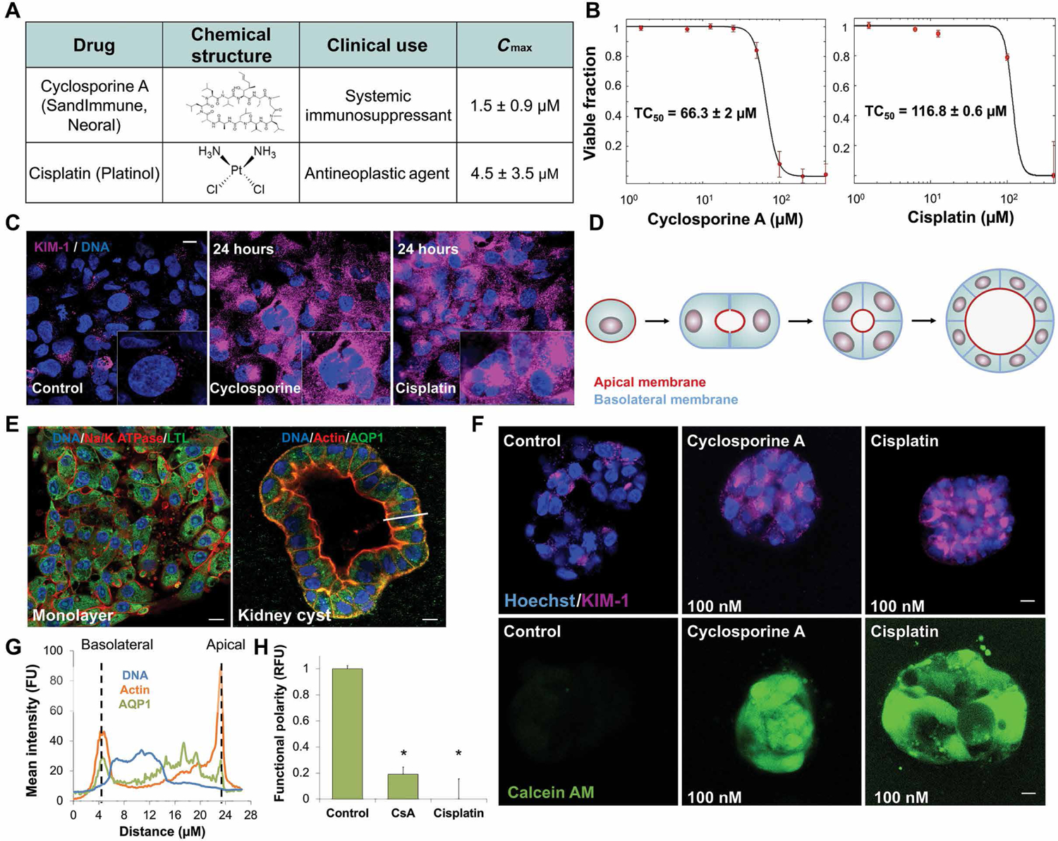
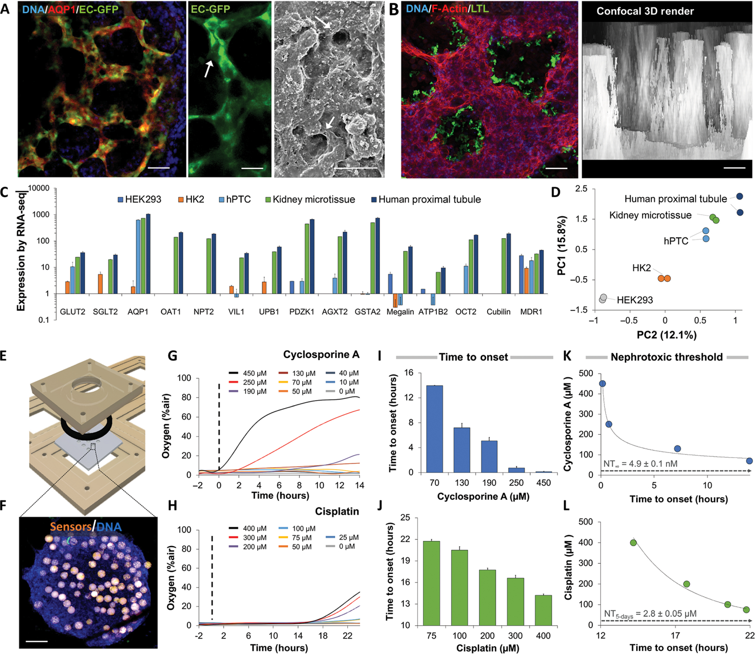
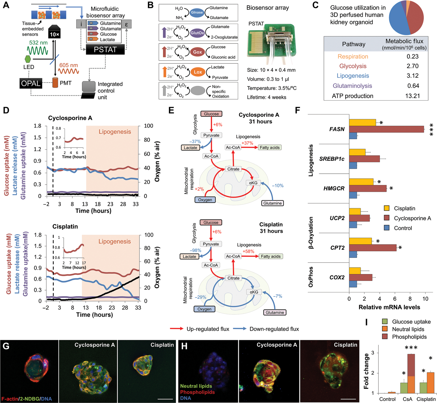
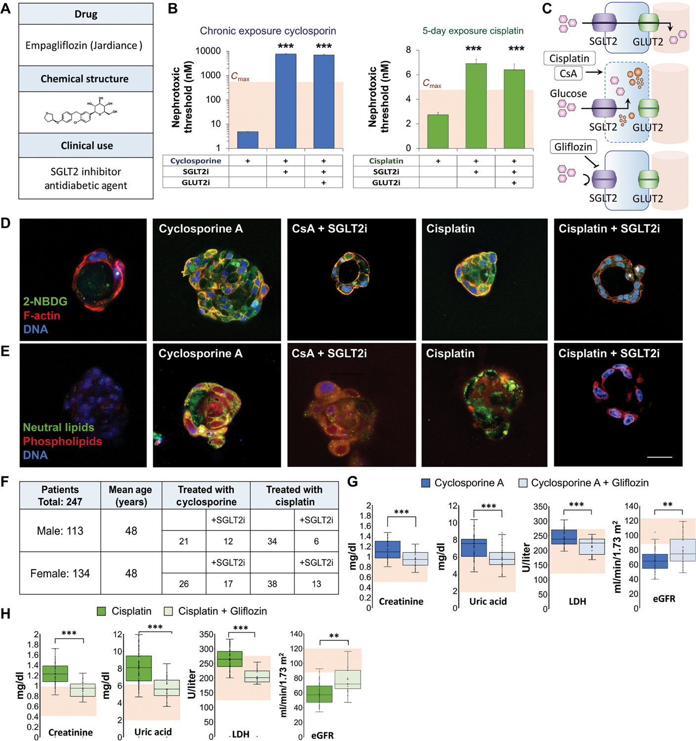
The kidney plays a critical role in fluid homeostasis, glucose control, and drug excretion. Loss of kidney function due to drug-induced nephrotoxicity affects over 20% of the adult population. The kidney proximal tubule is a complex vascularized structure that is particularly vulnerable to drug-induced nephrotoxicity. Here, we introduce a model of vascularized human kidney spheroids with integrated tissue-embedded microsensors for oxygen, glucose, lactate, and glutamine, providing real-time assessment of cellular metabolism. Our model shows that both the immunosuppressive drug cyclosporine and the anticancer drug cisplatin disrupt proximal tubule polarity at subtoxic concentrations, leading to glucose accumulation and lipotoxicity. Impeding glucose reabsorption using glucose transport inhibitors blocked cyclosporine and cisplatin toxicity by 1000- to 3-fold, respectively. Retrospective study of 247 patients who were diagnosed with kidney damage receiving cyclosporine or cisplatin in combination with the sodium-glucose cotransporter-2 (SGLT2) inhibitor empagliflozin showed significant (P < 0.001) improvement of kidney function, as well as reduction in creatinine and uric acid, markers of kidney damage. These results demonstrate the potential of sensor-integrated kidney-on-chip platforms to elucidate mechanisms of action and rapidly reformulate effective therapeutic solutions, increasing drug safety and reducing the cost of clinical and commercial failures.


Conflict of interest statement

Competing interests: Y.N. holds equity in Tissue Dynamics Ltd. Y.N., A.E., and A.C. are coinventors on international patent application number PCT/IL2020/050173, “A method for reduction of drug-induced nephrotoxicity,” associated with this study. The other authors declare that they have no competing interests.

Figures

Fig. 1.
Fig. 1.. Physiological doses of cyclosporine and cisplatin disrupt proximal tubule polarization.
(A) Table of structure, clinical use indications, and maximal physiological concentration (Cmax) of cyclosporine and cisplatin. (B) Dose-dependent toxicity curves of primary human proximal tubule cells (hPTCs) exposed for 24 hours to cyclosporine (TC50 = 66 μM) or cisplatin (TC50 = 95 μM) in 2D cell culture. (C) Immunofluorescence staining of acute kidney injury marker KIM-1 in hPTCs after 24-hour exposure to physiological doses of cyclosporine or cisplatin. (D) Schematic of 3D cyst formation, modeling proximal tubule polarity in vitro from single- and multicell suspensions in matrix. (E) Immunofluorescence staining of hPTCs in monolayer and 3D cyst formation. White bar indicates subsequent analyzed cross section in (G). (F) Functional polarity assay in cysts exposed to minute doses of cyclosporine or cisplatin for 24 hours. Fluorescent calcein is excreted through MDR1 (P-gp) in polarized cells. (G) Mean fluorescence intensity of cross section indicated by the white bar in (E), demonstrating polarized protein expression in hPTC cysts. FU, fluorescent unit; RFU, relative fluorescent unit. (H) Quantification of the loss of functional polarity. Scale bars, 10 μm. n = 4. N = 3. *P < 0.05, Student’s t test. Error bars indicate ± SE.
Fig. 2.
Fig. 2.. Characterization of nephrotoxicity in sensor-embedded vascularized kidney spheroids.
(A) Confocal microscopy of kidney spheroids composed of EGFP-labeled microvascular endothelial cells and aquaporin 1–positive (AQP1+) proximal tubule cells after 4 days in culture. Scanning electron micrograph of vascularized kidney spheroid shown on right. Arrows indicate patent endothelial lumens. Scale bars, 10 μm. (B) Confocal microscopy of proximal tubules expressing apical surface LTL in a spheroid. 3D reconstruction shows parallel longitudinal tubule-like structures 240 ± 40 μm in length. (C) Gene expression of RNA-seq data from human embryonic kidney (HEK) 293 cells, human kidney 2 (HK2) cells, and primary proximal tubule cells (hPTCs) in 2D culture compared to vascularized kidney spheroids and human proximal tubule tissue. (D) Principal component analysis (PCA) of gene expression patterns of vascularized kidney spheroids compared to HEK293, HK2, hPTCs, and proximal tubule tissue in vivo. (E) 3D design of a computer numerical control–fabricated nine-microwell bioreactor. Laser-cut disposable microwell chips containing nine spheroids are seeded in an open configuration and perfused until metabolic stabilization is achieved. (F) Fluorescent microscopy shows a human kidney spheroid with embedded oxygen sensors (orange). Scale bar, 100 μm. (G) Representative oxygen uptake over time response of kidney spheroids exposed to increasing concentrations of cyclosporine or (H) cisplatin. Dotted line notes onset of drug exposure. (I) Time to onset of response of kidney spheroids exposed to cyclosporine or (J) cisplatin. (K) Analytical derivation of nephrotoxic threshold (NT) using the time-to-onset–dependent flux accumulation equation. NT was defined as the horizontal asymptote, concentration for which the onset of damage will only occur at infinite time. Cyclosporine and (L) cisplatin showed NT of 4.9 ± 0.1 nM, a 1000-fold lower than clinically reported Cmax, and 2.8 ± 0.05 μM, respectively. Scale bars, 50 μm. N = 9. N = 3. Error bars indicate ± SE. For RNA-seq analysis, P values from a negative binomial Wald test are reported.
Fig. 3.
Fig. 3.. Metabolic analysis reveals drug-induced glucose accumulation and lipogenesis.
(A) Schematic of sensor-embedded spheroid-on-a-chip platform. The optical O2 measurement system (OPAL)–modulated light emitting diode (LED) signal excites tissue-embedded oxygen sensors. Phase shift is measured through a hardware-filtered photomultiplier (PMT). Chip outflow is connected to a biosensor array containing electrochemical sensors and an integrated potentiostat (PSTAT). Measurements are synchronized in real time by a single microprocessor. The arrows above the spheroids represent the direction of the flow. i, chemical input; ε, electrical output. (B) Photograph (right) and schematic (left) of microfluidic four-analyte biosensor array for glucose, lactate, glutamine, and glutamate. Glnase, glutaminase; GlutOx, l-glutamate oxidase; Gox, glucose oxidase; Lox, lactate oxidase. Array has an internal volume of 0.3 to 1 μl and integrated temperature control. (C) Intracellular metabolic fluxes of vascularized kidney spheroids in steady state. Glucose utilization and calculated ATP production are shown as nmol/min/106 cells. (D) Dynamics of oxygen, glucose, lactate, and glutamine fluxes during continuous exposure of vascularized kidney spheroids to physiological doses of cyclosporine or cisplatin under flow. n = 9. N = 3. (E) Flux balance analysis showing up-regulation of lipogenesis in kidney spheroids exposed to cyclosporine or cisplatin for 31 hours. Red and blue arrows note up- and down-regulated fluxes, respectively. (F) Relative gene expression after 48 hours of drug exposure. (G) Uptake and transport of 2-NBDG glucose analog in 3D hPTC cysts exposed to physiological concentrations of cyclosporine or cisplatin for 30 min. Student’s t test. n = 3. N = 3. (H) Fluorescence staining for neutral lipids and phospholipids in 3D cysts exposed to physiological concentrations of cyclosporine or cisplatin for 48 hours. (I) Quantification of glucose and lipid accumulation from (G) and (H). Scale bars, 25 μm. *P < 0.05, ***P < 0.001, Student’s t test. Error bars indicate ±SE. n = 4. N = 3.
Fig. 4.
Fig. 4.. Cyclosporine and cisplatin toxicity are reversed by SGLT2 inhibition.
(A) Structure and clinical indication of U.S. Food and Drug Administration–approved SGLT2 inhibitor (SGLT2i) empagliflozin. (B) Quantification of NTs in vascular kidney spheroids exposed to cyclosporin or cisplatin at various concentrations (400, 200, 100, 50, 25, 12, 6.2, 3.1, and 1.5 μM) in the presence of empagliflozin (SGLT2i) or empagliflozin and phloretin, glucose transporter-2 inhibitor (GLUT2i). Student’s t test. n = 9. N = 3. (C) Schematic of glucose transport in proximal tubule cells and mechanism of nephroprotective effect of empagliflozin (gliflozin). (D) Fluorescent glucose analog (2-NBDG) accumulation in 3D cysts exposed to low concentrations (100 nM) of cyclosporine or cisplatin for 30 min in the presence or absence of empagliflozin (SGLT2i). Control, cyclosporine A, and cisplatin images are reproduced from Fig. 3G for comparison. (E) Lipid accumulation in 3D cysts exposed to cyclosporine or cisplatin for 48 hours in the presence or absence of empagliflozin. Control, cyclosporine A, and cisplatin images are reproduced from Fig. 3H for comparison. (F) Table summarizing the number of patients with kidney damage in each group of a retrospective clinical study according to their treatment. (G) Box plots showing serum creatinine, uric acid, lactate dehydrogenase (LDH) concentrations, and estimated glomerular filtration rate (eGFR) in patients treated with cyclosporine compared to those treated with both cyclosporine and empagliflozin. Shaded area indicates normal values. (H) Box plots showing serum creatinine, uric acid, LDH concentrations, and eGFR in patients treated with cisplatin compared to those treated with both cisplatin and empagliflozin. Shaded area indicates normal values. Scale bars, 25 μm. Error bars indicate ±SE. Box plot center line indicates median; the cross indicates mean; box upper and lower limits indicate third and first quartiles, respectively; whiskers indicate 1.5× interquartile range; and points are data points. **P < 0.01, ***P < 0.001, Student’s t test.

Comment in

References

    1. Davis-Ajami ML, Fink JC, Wu J, Nephrotoxic medication exposure in U.S. Adults with predialysis chronic kidney disease: Health services utilization and cost outcomes. J. Manag. Care Spec. Pharm 22, 959–968 (2016). - PMC - PubMed
    1. Schetz M, Dasta J, Goldstein S, Golper T, Drug-induced acute kidney injury. Curr. Opin. Crit. Care 11, 555–565 (2005). - PubMed
    1. Perazella MA, Crystal-induced acute renal failure. Am. J. Med 106, 459–465 (1999). - PubMed
    1. Pisoni R, Ruggenenti P, Remuzzi G, Drug-induced thrombotic microangiopathy: Incidence, prevention and management. Drug Saf. 24, 491–501 (2001). - PubMed
    1. Medina PJ, Sipols JM, George JN, Drug-associated thrombotic thrombocytopenic purpura-hemolytic uremic syndrome. Curr. Opin. Hematol 8, 286–293 (2001). - PubMed

Publication types

Substances