. 2021 Apr 13;96(15):e1975-e1986.
doi: 10.1212/WNL.0000000000011745. Epub 2021 Feb 24.

Association of Memory Impairment With Concomitant Tau Pathology in Patients With Cerebral Amyloid Angiopathy

Affiliations

Association of Memory Impairment With Concomitant Tau Pathology in Patients With Cerebral Amyloid Angiopathy

Dorothee Schoemaker et al. Neurology. .

Abstract

Objective: Relying on tau-PET imaging, this cross-sectional study explored whether memory impairment is linked to the presence of concomitant tau pathology in individuals with cerebral amyloid angiopathy (CAA).

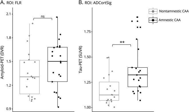
Methods: Forty-six patients with probable CAA underwent a neuropsychological examination and an MRI for quantification of structural markers of cerebral small vessel disease. A subset of these participants also completed a [11C]-Pittsburgh compound B (n = 39) and [18F]-flortaucipir (n = 40) PET for in vivo estimation of amyloid and tau burden, respectively. Participants were classified as amnestic or nonamnestic on the basis of neuropsychological performance. Statistical analyses were performed to examine differences in cognition, structural markers of cerebral small vessel disease, and amyloid- and tau-PET retention between participants with amnestic and those with nonamnestic CAA.

Results: Patients with probable CAA with an amnestic presentation displayed a globally more severe profile of cognitive impairment, smaller hippocampal volume (p < 0.001), and increased tau-PET binding in regions susceptible to Alzheimer disease neurodegeneration (p = 0.003) compared to their nonamnestic counterparts. Amnestic and nonamnestic patients with CAA did not differ on any other MRI markers or on amyloid-PET binding. In a generalized linear model including all evaluated neuroimaging markers, tau-PET retention (β = -0.85, p = 0.001) and hippocampal volume (β = 0.64 p = 0.01) were the only significant predictors of memory performance. The cognitive profile of patients with CAA with an elevated tau-PET retention was distinctly characterized by a significantly lower performance on the memory domain (p = 0.004).

Conclusions: These results suggest that the presence of objective memory impairment in patients with probable CAA could serve as a marker for underlying tau pathology.

Classification of evidence: This study provides Class II evidence that tau-PET retention is related to the presence of objective memory impairment in patients with CAA.


Figures

Figure 1
Figure 1. Flowchart Illustrating the Selection of Study Participants
Flowchart illustrating the selection of study participants from a larger study cohort (n = 78). Participants were classified as having probable cerebral amyloid angiopathy (CAA) or not on the basis of the modified Boston criteria., Three participants could not be classified due to an absence of research MRI and thus were excluded. MCI = mild cognitive impairment; PiB = Pittsburgh compound B.
Figure 2
Figure 2. Differences in Tau- and Amyloid-PET Retention Based on Amnestic Status in Individuals With CAA
Boxplots representing the dispersion of (A) amyloid-PET retention expressed as distribution volume ratio (DVR) in a region of interest (ROI) including the frontal, lateral temporal/parietal, and retrosplenial cortex (FLR) and (B) tau-PET retention expressed as standardized uptake value ratio (SUVR) in an ROI regrouping Alzheimer disease cortical signature regions (ADCortSig). Participants with probable cerebral amyloid angiopathy (CAA) with a nonamnestic presentation are represented in gray; those with an amnestic presentation are represented in black. ns = not statistically significant, **p < 0.01.
Figure 3
Figure 3. Associations Between Imaging Markers and Memory in Individuals With Probable CAA
Results of univariate generalized linear models between memory performance and (A) tau-PET retention expressed as standardized uptake value ratio (SUVR) in a region of interest regrouping Alzheimer disease cortical signature regions (ADCortSig) or (B) normalized total hippocampal volume (nHC). Dashed lines represent the 95% confidence intervals. (C) Bar graph representing the relative contribution of each evaluated regressor to the multivariate generalized linear model evaluating the contribution of neuroimaging markers on memory performance, using the LMG metric computed with the R package relaimpo (U. Grömping, 2006). Metrics are normalized to sum to 100%. Lines represent 95% confidence intervals after 1,000 bootstrapping replications. BPF = brain parenchymal fraction; CAA = cerebral amyloid angiopathy; CMB = cerebral microbleed; cSS = cortical superficial siderosis; DVR = distribution volume ratio; FLR = frontal, lateral temporal/parietal, and retrosplenial cortex; nWMH = normalized white matter hyperintensity volume. *p < 0.05; **p < 0.01.
Figure 4
Figure 4. Effect of Tau PET Status on the Cognitive Profile of Participants With Probable CAA
Bar graph contrasting the cognitive profile of participants with cerebral amyloid angiopathy (CAA) presenting with a positive or negative tau-PET. Tau-PET status was determined from the [18F]-flortaucipir standardized uptake value ratio in a region of interest comprising previously described Alzheimer disease cortical signature regions and using a previously established cutoff score. The p values were adjusted for multiple comparisons as per the Bonferroni correction. *Corrected p < 0.05; **corrected p < 0.01.

References

    1. Snyder HM, Corriveau RA, Craft S, et al. . Vascular contributions to cognitive impairment and dementia including Alzheimer's disease. Alzheimers Demen 2015;11:710–717. - PMC - PubMed
    1. De Reuck J. The impact of cerebral amyloid angiopathy in various neurodegenerative dementia syndromes: a neuropathological study. Neurol Res Int 2019;2019:7247325. - PMC - PubMed
    1. Boyle PA, Yu L, Wilson RS, Leurgans SE, Schneider JA, Bennett DA. Person‐specific contribution of neuropathologies to cognitive loss in old age. Ann Neurol 2018;83:74–83. - PMC - PubMed
    1. Thal DR, Griffin WST, de Vos RA, Ghebremedhin E. Cerebral amyloid angiopathy and its relationship to Alzheimer's disease. Acta Neuropathologica 2008;115:599–609. - PubMed
    1. Viswanathan A, Greenberg SM. Cerebral amyloid angiopathy in the elderly. Ann Neurol 2011;70:871–880. - PMC - PubMed

Publication types