

The Cytospora chrysosperma Virulence Effector CcCAP1 Mainly Localizes to the Plant Nucleus To Suppress Plant Immune Responses
- PMID: 33627507
- PMCID: PMC8544888
- DOI: 10.1128/mSphere.00883-20
The Cytospora chrysosperma Virulence Effector CcCAP1 Mainly Localizes to the Plant Nucleus To Suppress Plant Immune Responses
Abstract
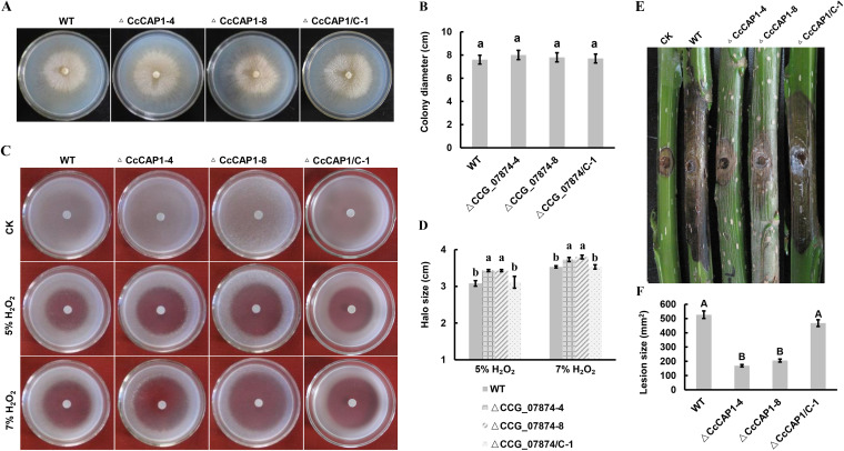
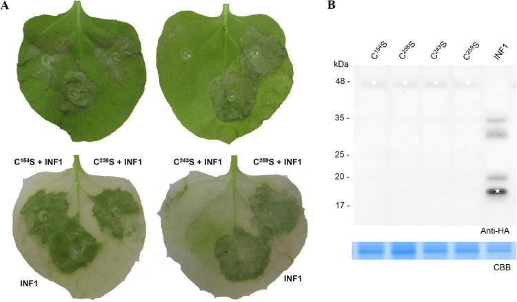
Canker disease is caused by the fungus Cytospora chrysosperma and damages a wide range of woody plants, causing major losses to crops and native plants. Plant pathogens secrete virulence-related effectors into host cells during infection to regulate plant immunity and promote colonization. However, the functions of C. chrysosperma effectors remain largely unknown. In this study, we used Agrobacterium tumefaciens-mediated transient expression system in Nicotiana benthamiana and confocal microscopy to investigate the immunoregulation roles and subcellular localization of CcCAP1, a virulence-related effector identified in C. chrysosperma CcCAP1 was significantly induced in the early stages of infection and contains cysteine-rich secretory proteins, antigen 5, and pathogenesis-related 1 proteins (CAP) superfamily domain with four cysteines. CcCAP1 suppressed the programmed cell death triggered by Bcl-2-associated X protein (BAX) and the elicitin infestin1 (INF1) in transient expression assays with Nicotiana benthamiana The CAP superfamily domain was sufficient for its cell death-inhibiting activity and three of the four cysteines in the CAP superfamily domain were indispensable for its activity. Pathogen challenge assays in N. benthamiana demonstrated that transient expression of CcCAP1 promoted Botrytis cinerea infection and restricted reactive oxygen species accumulation, callose deposition, and defense-related gene expression. In addition, expression of green fluorescent protein-labeled CcCAP1 in N. benthamiana showed that it localized to both the plant nucleus and the cytoplasm, but the nuclear localization was essential for its full immune inhibiting activity. These results suggest that this virulence-related effector of C. chrysosperma modulates plant immunity and functions mainly via its nuclear localization and the CAP domain.IMPORTANCE The data presented in this study provide a key resource for understanding the biology and molecular basis of necrotrophic pathogen responses to Nicotiana benthamiana resistance utilizing effector proteins, and CcCAP1 may be used in future studies to understand effector-triggered susceptibility processes in the Cytospora chrysosperma-poplar interaction system.
Keywords: Cytospora chrysosperma; plant immunity; subcellular localization; virulence effector.
Copyright © 2021 Han et al.
Figures


Similar articles
-
A Putative Effector CcSp84 of Cytospora chrysosperma Localizes to the Plant Nucleus to Trigger Plant Immunity.Int J Mol Sci. 2022 Jan 30;23(3):1614. doi: 10.3390/ijms23031614. Int J Mol Sci. 2022. PMID: 35163540 Free PMC article.
-
A putative elicitor CcHE1 from Cytospora chrysosperma enhances plant resistance to phytopathogenic fungi.Pest Manag Sci. 2025 Sep;81(9):5470-5483. doi: 10.1002/ps.8900. Epub 2025 May 13. Pest Manag Sci. 2025. PMID: 40357689
-
Trehalose Biosynthetic Genes Are Involved in the Development and Pathogenesis in the Poplar Canker Fungus Cytospora chrysosperma.Phytopathology. 2025 Mar;115(3):260-268. doi: 10.1094/PHYTO-05-24-0160-R. Epub 2025 Mar 22. Phytopathology. 2025. PMID: 39499502
-
Versatile effectors of phytopathogenic fungi target host immunity.J Integr Plant Biol. 2021 Nov;63(11):1856-1873. doi: 10.1111/jipb.13162. Epub 2021 Sep 15. J Integr Plant Biol. 2021. PMID: 34383388 Review.
-
Prediction of effector proteins and their implications in pathogenicity of phytopathogenic filamentous fungi: A review.Int J Biol Macromol. 2022 May 1;206:188-202. doi: 10.1016/j.ijbiomac.2022.02.133. Epub 2022 Feb 26. Int J Biol Macromol. 2022. PMID: 35227707 Review.
Cited by
-
Dual functionality of pathogenesis-related proteins: defensive role in plants versus immunosuppressive role in pathogens.Front Plant Sci. 2024 Aug 2;15:1368467. doi: 10.3389/fpls.2024.1368467. eCollection 2024. Front Plant Sci. 2024. PMID: 39157512 Free PMC article.
-
CcPmk1 is a regulator of pathogenicity in Cytospora chrysosperma and can be used as a potential target for disease control.Mol Plant Pathol. 2021 Jun;22(6):710-726. doi: 10.1111/mpp.13059. Epub 2021 Apr 9. Mol Plant Pathol. 2021. PMID: 33835616 Free PMC article.
-
Identification and Functional Analysis of CAP Genes from the Wheat Stripe Rust Fungus Puccinia striiformis f. sp. tritici.J Fungi (Basel). 2023 Jul 7;9(7):734. doi: 10.3390/jof9070734. J Fungi (Basel). 2023. PMID: 37504723 Free PMC article.
-
The mitogen-activated protein kinase module CcSte11-CcSte7-CcPmk1 regulates pathogenicity via the transcription factor CcSte12 in Cytospora chrysosperma.Stress Biol. 2024 Jan 16;4(1):4. doi: 10.1007/s44154-023-00142-w. Stress Biol. 2024. PMID: 38225467 Free PMC article.
-
Nuclear Effectors in Plant Pathogenic Fungi.Mycobiology. 2022 Sep 20;50(5):259-268. doi: 10.1080/12298093.2022.2118928. eCollection 2022. Mycobiology. 2022. PMID: 36404902 Free PMC article. Review.
References
-
- Kepley JB, Reeves FB, Jacobi WR, Adams GC. 2015. Species associated with Cytospora canker on Populus tremuloides. Mycotaxon 130:783–805. doi:10.5248/130.783. - DOI
-
- Adams GC, Roux J, Wingfield MJ. 2006. Cytospora species (Ascomycota, Diaporthales, and Valsaceae) introduced and native pathogens of trees in South Africa. Austral Plant Pathol 35:521–548. doi:10.1071/AP06058. - DOI
-
- Saitoh H, Fujisawa S, Mitsuoka C, Ito A, Hirabuchi A, Ikeda K, Irieda H, Yoshino K, Yoshida K, Matsumura H, Tosa Y, Win J, Kamoun S, Takano Y, Terauchi R. 2012. Large-scale gene disruption in Magnaporthe oryzae identifies MC69, a secreted protein required for infection by monocot and dicot fungal pathogens. PLoS Pathog 8:e1002711. doi:10.1371/journal.ppat.1002711. - DOI - PMC - PubMed
-
- Biggs AR, Davis DD, Merrill W. 1983. Histopathology of cankers on Populus caused by Cytospora chrysosperma. Can J Bot 61:563–574. doi:10.1139/b83-064. - DOI
Publication types
MeSH terms
Substances
Supplementary concepts
LinkOut - more resources
Full Text Sources
Other Literature Sources
Research Materials
Miscellaneous
