. 2021 Feb 24;11(1):4446.
doi: 10.1038/s41598-021-83869-8.

Thoracic shape changes in newborns due to their position

Affiliations

Thoracic shape changes in newborns due to their position

Serena de Gelidi et al. Sci Rep. .

Abstract

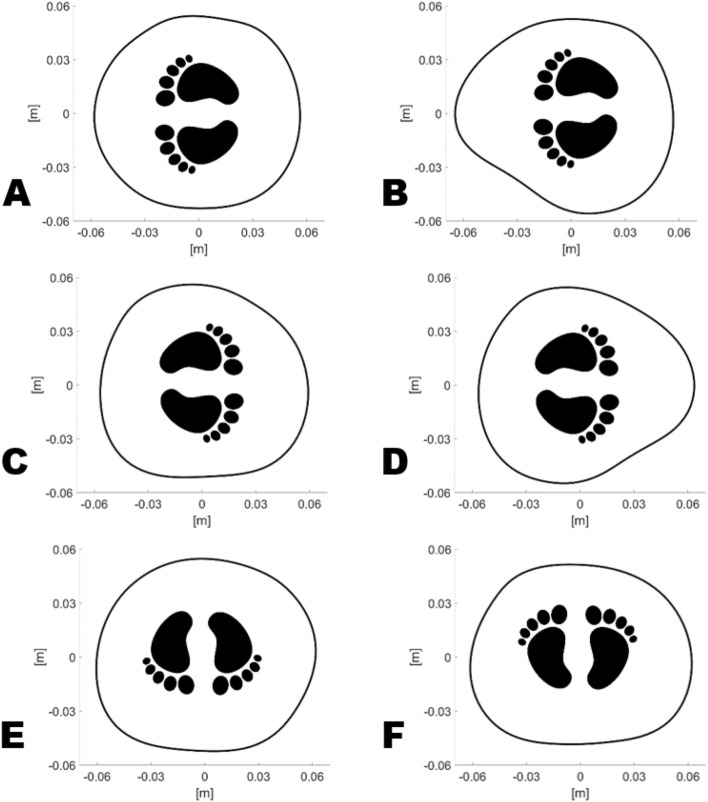
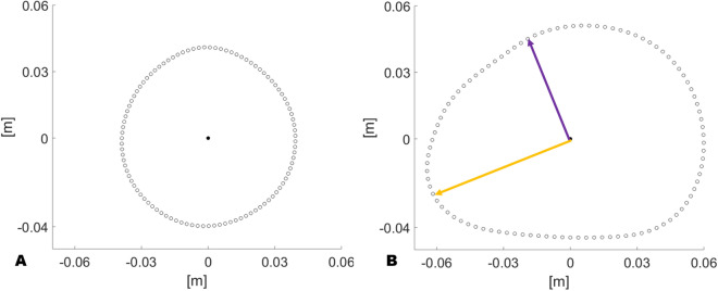
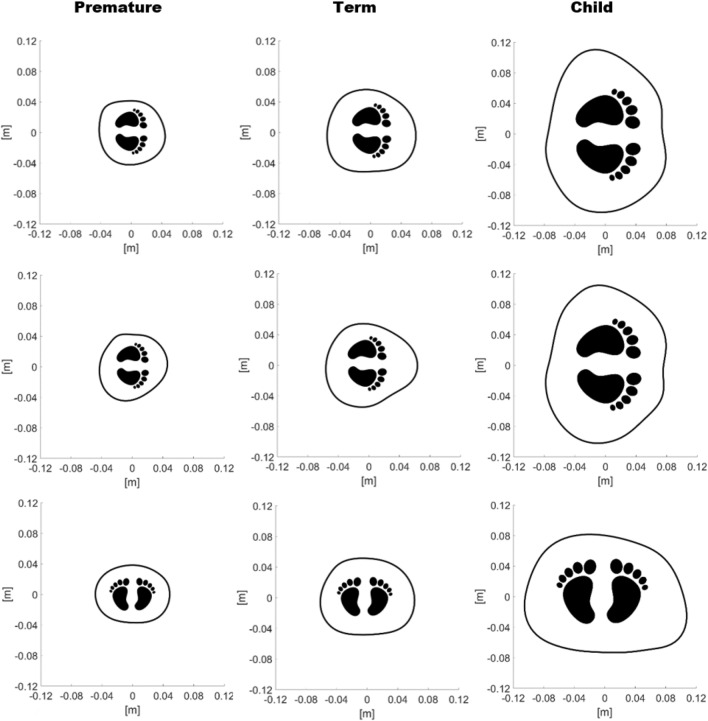
The highly compliant nature of the neonatal chest wall is known to clinicians. However, its morphological changes have never been characterized and are especially important for a customised monitoring of respiratory diseases. Here, we show that a device applied on newborns can trace their chest boundary without the use of radiation. Such technology, which is easy to sanitise between patients, works like a smart measurement tape drawing also a digital cross section of the chest. We also show that in neonates the supine position generates a significantly different cross section compared to the lateral ones. Lastly, an unprecedented comparison between a premature neonate and a child is reported.


Conflict of interest statement

The authors declare no competing interests.

Figures

Figure 1
Figure 1
The electronic measuring belt applied on one of the recruited subjects in the maternity ward. According to the protocol, in this shot the clinician was recording the torso shape of the baby lying on his left side with his arms up (A) and down (B). The photographs are published with permission of both of the baby’s parents.
Figure 2
Figure 2
Comparison of the computed chest boundaries for subject no. 30. The baby was lying on her right side with her arms up (A) and down (B), on her left with her arms up (C) and down (D), prone (E) and supine (F).
Figure 3
Figure 3
Comparison of the computed chest boundaries which feature the minimum (A) and the maximum (B) value of shape function. The purple arrow indicates the minimum distance from the centroid, whilst the orange one is the maximum distance.
Figure 4
Figure 4
Evolution of the torso boundary computed for patient no. 31 (premature), patient no. 30 (term) and a child. On the top row the subjects are lying on their left side without compressing the torso with their arm. On the middle row their left arm lies between the mattress and the chest. On the bottom row the study subjects are lying supine.
Figure 5
Figure 5
Algorithm steps (green) allowed to double check the data acquisition during the protocol, which was carried out by a clinician (red) supported by an engineer (blue). A previous work explains the core of the algorithm.

Similar articles

Cited by

References

    1. WHO . Preterm Birth. Geneva: WHO; 2018.
    1. Sophocleous L, et al. Clinical performance of a novel textile interface for neonatal chest electrical impedance tomography. Physiol. Meas. 2018 doi: 10.1088/1361-6579/aab513. - DOI - PubMed
    1. Rahtu M, et al. Early recognition of pneumothorax in neonatal respiratory distress syndrome with electrical impedance tomography. Am. J. Respir. Crit. Care Med. 2019;200:1060–1061. doi: 10.1164/rccm.201810-1999IM. - DOI - PubMed
    1. Kallio M, et al. Electrical impedance tomography reveals pathophysiology of neonatal pneumothorax during NAVA. Clin. Case Rep. 2020;8:1574–1578. doi: 10.1002/ccr3.2944. - DOI - PMC - PubMed
    1. Stocks J, Hislop A. Structure and function of the respiratory system: Developmental aspects and their relevance to aerosol therapy. In: Bisgaard H, O’Callaghan C, Smaldone G, editors. Drug Delivery to the Lung. New York: Taylor & Francis Group; 2001.

Publication types