Review
. 2021 Feb;53(2):202-209.
doi: 10.1038/s12276-021-00568-0. Epub 2021 Feb 24.

Transcriptional regulatory network for the establishment of CD8+ T cell exhaustion

Affiliations
Review

Transcriptional regulatory network for the establishment of CD8+ T cell exhaustion

Wooseok Seo et al. Exp Mol Med. 2021 Feb.

Abstract

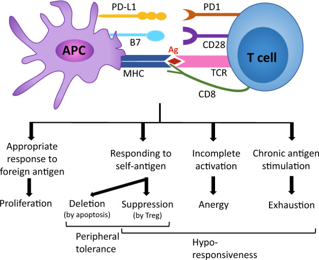
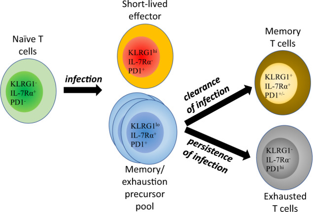
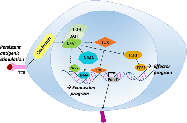
Chronic infection with persistent antigenic stimulation results in the generation of exhausted CD8+ T cells, which are considered defective effector CD8+ T cells, and thus compromises effective immune responses. However, recent studies have illustrated that exhausted CD8+ T cells may be purposely generated and maintained to provide mild immune responses against chronic infection or cancer, which can be safer over a long period of time than strong immune responses. Indeed, a specific population of exhausted CD8+ T cells that behaves similarly to self-renewing stem cells and provides a continuous supply of exhausted CD8+ T cells has been identified, indicating that this population can be considered progenitors of exhausted CD8+ T cells. Furthermore, several ground-breaking studies in the last few years have shed new light on the transcriptional regulatory network governing the generation and propagation of exhausted CD8+ T cells, which involves T cell receptor (TCR) signaling that leads to NFAT-TCF1 (nuclear factor of activated T cells-T cell factor 1) activity followed by activation of the TOX/NR4A axis. Elucidation of the intracellular signaling pathways will help to define the definitive developmental stages leading to exhausted CD8+ T cells, which can be exploited to advance our never-ending battle against cancer. This review will summarize the recent discoveries that have deepened our understanding of the exhaustion program of cytotoxic CD8+ T cells.


Conflict of interest statement

H.N. received honoraria and research funding from Ono Pharmaceutical, Chugai Pharmaceutical, MSD and Bristol-Myers Squibb and research funding from Taiho Pharmaceutical, Daiichi-Sankyo, Kyowa Kirin, Zenyaku Kogyo, Oncolys BioPharma, Debiopharma, Asahi-Kasei, Sysmex, Fujifilm, SRL, Astellas Pharmaceutical, Sumitomo Dainippon Pharma and BD Japan outside of this study. The other authors declare no competing interests.

Figures

Fig. 1
Fig. 1. Hyporesponsiveness of CD8+ T cells.
Naïve cytotoxic CD8+ T cells scan the cell surface of antigen-presenting cells (APCs), such as dendritic cells (DCs), which present processed peptides on MHC molecules. The inputs from T cell receptors (TCRs) function as signal 1, those from costimulatory molecules (such as CD28, OX40, and ICOS) act as signal 2, and cytokines (such as IL-12) provide signal 3; these signals are combined to generate appropriate intracellular signaling events that dictate the outcome of T cell fate. Recognition of cognate foreign antigens with appropriate signals 2 and 3 activates CD8+ T cells to differentiate into effector T cells. CD8+ T cells responding to self-antigens are subjected to peripheral tolerance, in which they are either deleted by apoptosis or inactivated (or suppressed) by regulatory T cells (Tregs). Insufficient signals 2 and 3 or strong negative signals provided by coinhibitory molecules (such as PD1 and CTLA4) result in incomplete activation of T cells, which are then called anergic T cells. Persistent cognate antigen stimulation during chronic infection or cancer induces another hyporesponsive state of T cells called exhaustion, which is characterized by high expression of multiple coinhibitory molecules.
Fig. 2
Fig. 2. Differentiation of exhausted T cells.
T cell activation generates both short-lived effector cells with terminally differentiated features and memory precursors. If the infection is cleared without delay (acute infection), the short-lived effector cells die, and the memory precursors give rise to long-lived memory T cells. However, if the infection persists without resolution (chronic infection), the memory precursors generate PD1+CXCR5+TCF1+ progenitors of exhausted T cells that self-renew to maintain the pool of mature exhausted T cells (PD1+TCF1-). It appears that these progenitors of exhausted T cells phenotypically resemble memory progenitors during acute infection, but they functionally differ in the generation of daughter cells.
Fig. 3
Fig. 3. Transcriptional regulatory network of exhausted CD8+ T cells.
T cell receptor (TCR) activation triggers phosphorylation of the kinase Lck, which in turn activates calcineurin pathways. Calcineurin dephosphorylates cytoplasmic nuclear factor of activated T cells (NFAT), which then translocates into the nucleus and regulates genes involved in either T cell effector function or T cell exhaustion. Therefore, the transduction of extracellular signals by NFAT is one of the most important steps in T cell exhaustion. On the one hand, NFAT represses TCF1 in exhausted CD8+ T cells since TCF1 can induce the formation of terminal effector and memory CD8+ T cells by upregulating EOMES expression. On the other hand, NFAT induces the transcription factors TOX and NR4A, which execute the exhaustion program of CD8+ T cells. It should be noted that TCF1 is necessary in the progenitors of exhausted T cells, but it should be repressed in terminally differentiated exhausted T cells.

References

    1. Taniuchi I. CD4 helper and CD8 cytotoxic T cell differentiation. Annu. Rev. Immunol. 2018;36:579–601. - PubMed
    1. Sprent J, Surh CD. Normal T cell homeostasis: the conversion of naive cells into memory-phenotype cells. Nat. Immunol. 2011;12:478–484. - PMC - PubMed
    1. Neefjes J, Jongsma ML, Paul P, Bakke O. Towards a systems understanding of MHC class I and MHC class II antigen presentation. Nat. Rev. Immunol. 2011;11:823–836. - PubMed
    1. Smith-Garvin JE, Koretzky GA, Jordan MS. T cell activation. Annu. Rev. Immunol. 2009;27:591–619. - PMC - PubMed
    1. Joshi NS, et al. Inflammation directs memory precursor and short-lived effector CD8(+) T cell fates via the graded expression of T-bet transcription factor. Immunity. 2007;27:281–295. - PMC - PubMed

Publication types

MeSH terms