Review
. 2021 Aug;18(8):565-580.
doi: 10.1038/s41569-021-00517-4. Epub 2021 Feb 24.

The quiescent endothelium: signalling pathways regulating organ-specific endothelial normalcy

Affiliations
Review

The quiescent endothelium: signalling pathways regulating organ-specific endothelial normalcy

Nicolas Ricard et al. Nat Rev Cardiol. 2021 Aug.

Abstract

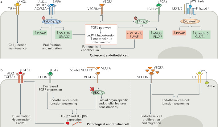
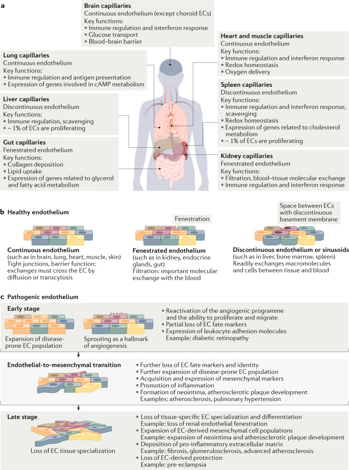
Endothelial cells are at the interface between circulating blood and tissues. This position confers on them a crucial role in controlling oxygen and nutrient exchange and cellular trafficking between blood and the perfused organs. The endothelium adopts a structure that is specific to the needs and function of each tissue and organ and is subject to tissue-specific signalling input. In adults, endothelial cells are quiescent, meaning that they are not proliferating. Quiescence was considered to be a state in which endothelial cells are not stimulated but are instead slumbering and awaiting activating signals. However, new evidence shows that quiescent endothelium is fully awake, that it constantly receives and initiates functionally important signalling inputs and that this state is actively regulated. Signalling pathways involved in the maintenance of functionally quiescent endothelia are starting to be identified and are a combination of endocrine, autocrine, paracrine and mechanical inputs. The paracrine pathways confer a microenvironment on the endothelial cells that is specific to the perfused organs and tissues. In this Review, we present the current knowledge of organ-specific signalling pathways involved in the maintenance of endothelial quiescence and the pathologies associated with their disruption. Linking organ-specific pathways and human vascular pathologies will pave the way towards the development of innovative preventive strategies and the identification of new therapeutic targets.


Conflict of interest statement

The authors declare no competing interests.

Figures

Fig. 1
Fig. 1. Tissue specificity of signalling pathways implicated in endothelial quiescence.
Quiescent endothelial cells receive autocrine, endocrine and paracrine signalling inputs. Paracrine stimulation confers a tissue-specific microenvironment to the endothelium. For example, vascular endothelial growth factor (VEGF) signalling is crucial to the formation and maintenance of fenestrated endothelia, whereas paracrine WNT signalling induces formation and maintenance of continuous endothelial lining, which is crucial for vascular integrity in general and that of the blood–brain barrier in particular. Autocrine transforming growth factor-β (TGFβ) signalling is a signature of endothelial cell dysfunction. ANG1, angiopoietin 1; ACVR2A, activin A receptor type 2A; ALK, activin receptor-like kinase; BMP9, bone morphogenetic protein 9; BMPR2, bone morphogenetic protein receptor type 2; FGF, fibroblast growth factor; FGFR, fibroblast growth factor receptor; LRP, lipoprotein receptor-related protein; TGFβR, transforming growth factor-β receptor; TIE2, angiopoietin 1 receptor; VEGFR, vascular endothelial growth factor receptor.
Fig. 2
Fig. 2. Signalling crosstalk in quiescent and activated endothelial cells.
a | Regulation of endothelial fenestration is achieved by a combination of signalling pathways regulating Plvap expression. Inhibition of the transforming growth factor-β (TGFβ) signalling is controlled by several signalling pathways including bone morphogenetic protein (BMP), fibroblast growth factor (FGF) and vascular endothelial growth factor (VEGF) signalling circuits. b | Pathological endothelium. Decreased FGF or VEGF signalling input leads to activation of an autocrine TGFβ signalling loop that, in turn, induces inflammation, hypertension and endothelial-to-mesenchymal transition (EndMT). Excessive VEGF signalling and autocrine angiopoietin 2 (ANG2) signalling induce pathological angiogenesis. ACVR2A, activin A receptor type 2A; ALK, activin receptor-like kinase; BMPR2, bone morphogenetic protein receptor type 2; eNOS, endothelial nitric oxide synthase; ERK, extracellular signal-regulated kinase; FGFR, fibroblast growth factor receptor; GLUT1, glucose transporter 1; LRP, lipoprotein receptor-related protein; PLVAP, plasmalemma vesicle-associated protein; SMAD, mothers against decapentaplegic homologue; TIE2, angiopoietin 1 receptor; VEGFR, vascular endothelial growth factor receptor.
Fig. 3
Fig. 3. Endothelial heterogeneity in health and disease.
a | Quiescent endothelial cell (EC) heterogeneity in structure, function, immune regulation (interferon response and leukocyte adhesion molecule expression) and metabolism between tissues and within tissues. The information shown in this panel a is from ref.. b | Heterogeneity in healthy capillary ECs between organs. c | Development of endothelial dysfunction. This is a stepwise process, progressing from activation of ECs to the development of endothelial-to-mesenchymal transition to the full-blown pathological end state. This sequence of events leads to ECs losing their normal fate and acquiring features of mesenchymal cell types, including fibroblasts, smooth muscle cells and macrophages, in a process known as endothelial-to-mesenchymal transition. These events result in the initiation and propagation of inflammation, loss of normal endothelial structures and function, increased vascular permeability and formation of pathological lesions, such as atherosclerotic plaques.

References

    1. Al-Soudi A, Kaaij MH, Tas SW. Endothelial cells: from innocent bystanders to active participants in immune responses. Autoimmun. Rev. 2017;16:951–962. doi: 10.1016/j.autrev.2017.07.008. - DOI - PubMed
    1. Yau JW, Teoh H, Verma S. Endothelial cell control of thrombosis. BMC Cardiovasc. Disord. 2015;15:130. doi: 10.1186/s12872-015-0124-z. - DOI - PMC - PubMed
    1. Konukoglu D, Uzun H. Endothelial dysfunction and hypertension. Adv. Exp. Med. Biol. 2017;956:511–540. doi: 10.1007/5584_2016_90. - DOI - PubMed
    1. Komarova YA, Kruse K, Mehta D, Malik AB. Protein interactions at endothelial junctions and signaling mechanisms regulating endothelial permeability. Circ. Res. 2017;120:179–206. doi: 10.1161/CIRCRESAHA.116.306534. - DOI - PMC - PubMed
    1. Chen W, et al. The endothelial tip-stalk cell selection and shuffling during angiogenesis. J. Cell Commun. Signal. 2019;13:291–301. doi: 10.1007/s12079-019-00511-z. - DOI - PMC - PubMed

Publication types