. 2021 Feb 8:12:601404.
doi: 10.3389/fphar.2021.601404. eCollection 2021.

Reduction of Neuroinflammation by δ-Opioids Via STAT3-Dependent Pathway in Chronic Glaucoma Model

Affiliations

Reduction of Neuroinflammation by δ-Opioids Via STAT3-Dependent Pathway in Chronic Glaucoma Model

Shahid Husain et al. Front Pharmacol. .

Abstract

The main objective of this study was to determine the inhibition of pro-inflammatory cytokines and their associated signaling molecules by δ-opioid receptor activation by a selective ligand, SNC-121 in chronic rat glaucoma model. Intraocular pressure was raised in rat eyes by injecting 2 M hypertonic saline into the limbal veins. SNC-121 (1 mg/kg; i. p) or Stattic (5 mg/kg; i. p) was administered in Brown Norway rats daily for 7 days. The mRNA expression of IL-1β, TNF-α, Fas, IL-6, leukemia inhibitory factor, and IFN-γ was increased significantly in the retina of ocular hypertensive animals at day 7, post injury. Administration of SNC-121 (1 mg/kg; i. p. injection) for 7 days (once a day) completely inhibited the increase in the mRNA and protein expression of pro-inflammatory cytokines. Mechanistically, we provide data showing a significant increase in the phosphorylation of STAT3 at tyrosine 705 whereas a moderate but significant increase in the total STAT3 protein expression was also seen in the retina of ocular hypertensive animals. Data illustrated that SNC-121 administration completely abrogated ocular hypertension-induced increase in STAT3Y705 phosphorylation. Interestingly, acetylation of STAT3 at lysine 685 (AcK685) was reduced in ocular hypertensive animals and subsequently increased significantly by SNC-121 treatment. Stattic, a selective STAT3 inhibitor, administration resulted in a complete attenuation in the production of IL-1β and IL-6 in ocular hypertensive animals. In conclusion, δ-opioid receptor activation suppressed the phosphorylation of STAT3 at tyrosine 705 and increased acetylation at lysine 686 and these posttranslational modifications can regulate the production of some but not all pro-inflammatory cytokines in response to glaucomatous injury.

Keywords: glaucoma; neuroinflammantion; opioids; protein acetylation; transcription factors.


Conflict of interest statement

The authors declare that the research was conducted in the absence of any commercial or financial relationships that could be construed as a potential conflict of interest.

Figures

FIGURE 1
FIGURE 1
Changes in mRNA expression of pro-inflammatory cytokines in response to ocular hypertension and SNC-121 treatment. Intraocular pressure (IOP) was raised by 2.0 M hypertonic saline injection followed by δ-opioid receptor agonist (1 mg/kg; i. p injections) treatment for 7 days, once a day. The retinas were collected at day 7, post hypertonic saline injection and mRNA was analyzed for IL-1β (A), TNF-α (B), FAS (C), IL-6 (D), LIF (E), and IFN-γ (F) using cDNA that was synthesized from 1 µg total RNA. The relative changes in mRNA levels were measured by quantitative RT-PCR (qRT-PCR) using primers specific for each gene as indicated in Table 1. The qRT-PCR data was normalized using ß-actin gene expression as an internal control. The relative expression was calculated based on the comparative threshold cycle (2−ΔΔCt) method. Bar graph represents mean data ±SEM. OH Eyes; ocular hypertensive eyes; OH Eyes + SNC-121, ocular hypertensive eyes with SNC-121 treatment. *p < 0.05; **p < 0.01; ***p < 0.001; ****p < 0.0001; n = 4–9. In this experiment “n” represent a biological replicate.
FIGURE 2
FIGURE 2
Effects of δ-opioid receptor agonist (SNC-121) treatment on interleukin-1β (IL-1β) (A) and interleukin-6 (IL-6) (B) production in ocular hypertensive animal. Animals were euthanized and eyes were enucleated at day 7, post hypertonic saline injections. Retina cryosections were immunostained using a selective anti-IL-1β or anti-IL-6 antibody. The nuclei were counterstained with DAPI. Red color indicates staining for IL-1β and IL-6 and blue for the nuclei. There was no positive immunostaining when primary antibodies were omitted (not shown). Data shown here is a representation of at least four independent experiments. We have used four animals in each treatment group. OH Eyes; ocular hypertensive eyes; OH Eyes + SNC-121, ocular hypertensive eyes with SNC-121 treatment, bar size 20 µm.
FIGURE 3
FIGURE 3
Effects of SNC-121 treatment on mRNA expression of STAT1 (A), STAT2 (B), and STAT3 (C). The retina from normal, ocular hypertensive, and SNC-121 treated ocular hypertensive animals were collected at day 7, post hypertonic saline injection. Complementary DNA was synthesized from 1 µg total RNA extracted from retina. The relative changes in mRNA levels were measured by quantitative RT-PCR (qRT-PCR) using primers specific for each gene as indicated in Table 1. The qRT-PCR data was normalized using ß-actin gene expression as an internal control. The relative expression was calculated based on the comparative threshold cycle (2−ΔΔCt) method. Bar graph represents mean data ±SEM. OH Eyes; ocular hypertensive eyes; OH Eyes + SNC-121, ocular hypertensive eyes with SNC-121 treatment. **p < 0.01; ***p < 0.001; n = 4–9. In this experiment “n” represent a biological replicate.
FIGURE 4
FIGURE 4
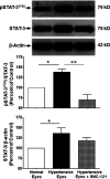
Changes in the phosphorylation of STAT3 (Tyr 705) and total STAT3 by SNC-121 treatment in ocular hypertensive animals. The retina from normal, ocular hypertensive, and SNC-121 treated ocular hypertensive animals were collected at day 7, post hypertonic saline injection. Equivalent amount of retina protein extracts (20 μg) were analyzed by Western blotting using a selective anti-phosphoSTAT3 (Tyr 705), anti-STAT3, and anti-β-actin antibodies. The band intensities were measured using enhanced chemiluminescence reagent and Versadoc imaging system. Bar graph represents mean data ±SEM. OH Eyes; ocular hypertensive eyes; OH Eyes + SNC-121, ocular hypertensive eyes with SNC-121 treatment. *p < 0.05; ****p < 0.0001; n = 6–9. In this experiment “n” represent a biological replicate.
FIGURE 5
FIGURE 5
Changes in the signal transducer and activator of transcription 3 (STAT3) phosphorylation in response to ocular hypertension and δ-opioid receptor agonist (SNC-121) treatment. The retina from normal, ocular hypertensive, and SNC-121 treated ocular hypertensive animals were collected at day 7, post hypertonic saline injection. Retina cryosections were immunostained using a selective anti-phosphoSTAT3 (Tyr 705) antibody and the nuclei were counterstained with DAPI. Green color indicates staining for phosphorylation of STAT3 and blue for the nuclei. There was no positive immunostaining when primary antibodies were omitted (not shown). Data shown here is a representation of at least four independent experiments. We have used four animals in each treatment group. OH Eyes; ocular hypertensive eyes; OH Eyes + SNC-121, ocular hypertensive eyes with SNC-121 treatment, bar size 20 µm.
FIGURE 6
FIGURE 6
Changes in STAT3 acetylation at lysine 685 (AcK685) in response to ocular hypertension and SNC-121 treatment. The retina from normal, ocular hypertensive, and SNC-121 treated ocular hypertensive animals were collected at day 7, post hypertonic saline injection. Equivalent amount of retina protein extracts (20 μg) were analyzed by Western blotting using a selective anti-Acetyl STAT3 (AcK685) or anti-β-actin antibodies. The band intensities were measured using enhanced chemiluminescence reagent and Versadoc imaging system. Bar graph represents mean data ±SEM. OH Eyes; ocular hypertensive eyes; OH Eyes + SNC-121, ocular hypertensive eyes with SNC-121 treatment. *p < 0.05. n = 6, In this experiment “n” represent a biological replicate.
FIGURE 7
FIGURE 7
Inhibitory effects of Stattic, a selective STAT3 inhibitor, on the mRNA expression of IL-1β (A), TNF-α (B), IL-6 (C), and IFN-γ (D). The retina from normal, ocular hypertensive, and Stattic treated ocular hypertensive animals were collected at day 7, post hypertonic saline injection. Complementary DNA was synthesized from 1 µg total RNA extracted from retina. The relative changes in mRNA levels were measured by quantitative RT-PCR (qRT-PCR) using primers specific for each gene as indicated in Table 1. The qRT-PCR data was normalized using ß-actin gene expression as an internal control. The relative expression was calculated based on the comparative threshold cycle (2−ΔΔCt) method. Bar graph represents mean data ±SEM. OH Eyes; ocular hypertensive eyes; OH Eyes + Stattic, ocular hypertensive eyes with Stattic treatment. *p < 0.05; **p < 0.01; ***p < 0.001; n = 6–9. In this experiment “n” represent a biological replicate.

Similar articles

Cited by

References

    1. Abdul Y., Akhter N., Husain S. (2013). Delta-opioid agonist SNC-121 protects retinal ganglion cell function in a chronic ocular hypertensive rat model. Invest. Ophthalmol. Vis. Sci. 54 (3), 1816–1828. 10.1167/iovs.12-10741 - DOI - PMC - PubMed
    1. Adornetto A., Russo R., Parisi V. (2019). Neuroinflammation as a target for glaucoma therapy. Neural Regen Res. 14 (3), 391–394. 10.4103/1673-5374.245465 - DOI - PMC - PubMed
    1. Airaksinen P. J., Heijl A. (1983). Visual field and retinal nerve fibre layer in early glaucoma after optic disc haemorrhage. Acta Ophthalmol. 61 (2), 186–194. 10.1111/j.1755-3768.1983.tb01412.x - DOI - PubMed
    1. Akhter N., Nix M., Abdul Y., Singh S., Husain S. (2013a). Delta-opioid receptors attenuate TNF-alpha-induced MMP-2 secretion from human ONH astrocytes. Invest. Ophthalmol. Vis. Sci. 54 (10), 6605–6611. 10.1167/iovs.13-12196 - DOI - PMC - PubMed
    1. Akhter N., Nix M., Abdul Y., Singh S., Husain S. (2013b). Delta-opioid receptors attenuate TNF-α-induced MMP-2 secretion from human ONH astrocytes. Invest. Ophthalmol. Vis. Sci. 54 (10), 6605–6611. 10.1167/iovs.13-12196 - DOI - PMC - PubMed

LinkOut - more resources