

Isoegomaketone from Perilla frutescens (L.) Britt Stimulates MAPK/ERK Pathway in Human Keratinocyte to Promote Skin Wound Healing
- PMID: 33628306
- PMCID: PMC7889401
- DOI: 10.1155/2021/6642606
Isoegomaketone from Perilla frutescens (L.) Britt Stimulates MAPK/ERK Pathway in Human Keratinocyte to Promote Skin Wound Healing
Abstract
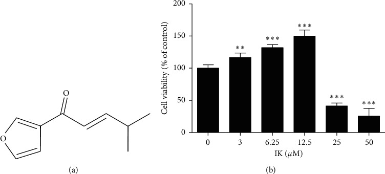
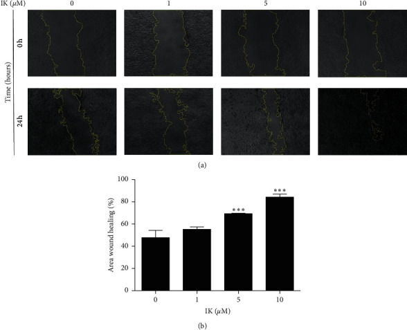
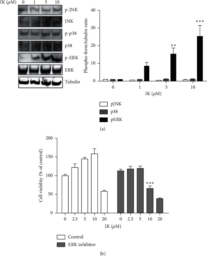
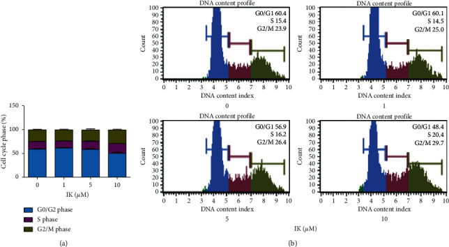
Skin wound healing is essential for recovery from injury, and delayed or impaired wound healing is a severe therapeutic challenge. Keratinocytes, a major component of the epidermis, play crucial roles in reepithelialization during wound healing including cell proliferation. Recent studies have shown that compounds from natural products have candidates for healing skin injury. Isoegomaketone (IK), isolated from leaves of Perilla frutescens var. crispa (Lamiaceae), has various bioactivities. However, the effect of IK on cutaneous wound healing processes has not been studied yet. In this study, we demonstrated that IK exhibits therapeutic wound healing effects using the human keratinocyte cell line HaCaT. Notably, IK promoted cell proliferation and migration in a dose-dependent manner in vitro, and treatment with 10 μM IK upregulated these processes by approximately 1.5-fold after 24 h compared with the control. IK induced the activation of the MAPK/ERK pathway and cell cycle progression to the S and G2/M phases. Thus, this study demonstrates IK as a potential candidate to upregulate wound healing that may provide therapeutic benefits to patients with delayed wound healing.
Copyright © 2021 Ye-Ram Kim et al.
Conflict of interest statement
The authors declare no conflicts of interest.
Figures
Similar articles
-
Advances in the Pharmacological Activities and Effects of Perilla Ketone and Isoegomaketone.Evid Based Complement Alternat Med. 2022 Oct 28;2022:8809792. doi: 10.1155/2022/8809792. eCollection 2022. Evid Based Complement Alternat Med. 2022. PMID: 36337585 Free PMC article. Review.
-
Isoegomaketone Upregulates Heme Oxygenase-1 in RAW264.7 Cells via ROS/p38 MAPK/Nrf2 Pathway.Biomol Ther (Seoul). 2016 Sep 1;24(5):510-6. doi: 10.4062/biomolther.2015.194. Biomol Ther (Seoul). 2016. PMID: 27582555 Free PMC article.
-
Isoegomaketone inhibits lipopolysaccharide-induced nitric oxide production in RAW 264.7 macrophages through the heme oxygenase-1 induction and inhibition of the interferon-beta-STAT-1 pathway.J Agric Food Chem. 2010 Jan 27;58(2):860-7. doi: 10.1021/jf9033333. J Agric Food Chem. 2010. PMID: 20030328
-
A New Monoterpene from the Leaves of a Radiation Mutant Cultivar of Perilla frutescens var. crispa with Inhibitory Activity on LPS-Induced NO Production.Molecules. 2017 Sep 4;22(9):1471. doi: 10.3390/molecules22091471. Molecules. 2017. PMID: 28869556 Free PMC article.
-
Wound Healing and the Use of Medicinal Plants.Evid Based Complement Alternat Med. 2019 Sep 22;2019:2684108. doi: 10.1155/2019/2684108. eCollection 2019. Evid Based Complement Alternat Med. 2019. PMID: 31662773 Free PMC article. Review.
Cited by
-
Defining the Potential Targets for Biological Activity of Isoegomaketone Based on Network Pharmacology and Molecular Docking Methods.Life (Basel). 2022 Dec 15;12(12):2115. doi: 10.3390/life12122115. Life (Basel). 2022. PMID: 36556480 Free PMC article.
-
Effect of Electrical Stimulation on Diabetic Human Skin Fibroblast Growth and the Secretion of Cytokines and Growth Factors Involved in Wound Healing.Biology (Basel). 2021 Jul 9;10(7):641. doi: 10.3390/biology10070641. Biology (Basel). 2021. PMID: 34356496 Free PMC article.
-
A frog peptide provides new strategies for the intervention against skin wound healing.Cell Mol Biol Lett. 2023 Jul 28;28(1):61. doi: 10.1186/s11658-023-00468-3. Cell Mol Biol Lett. 2023. PMID: 37501100 Free PMC article.
-
Advances in the Pharmacological Activities and Effects of Perilla Ketone and Isoegomaketone.Evid Based Complement Alternat Med. 2022 Oct 28;2022:8809792. doi: 10.1155/2022/8809792. eCollection 2022. Evid Based Complement Alternat Med. 2022. PMID: 36337585 Free PMC article. Review.
-
Efficacy of Perilla frutescens (L.) Britton var. frutescens extract on mild knee joint pain: A randomized controlled trial.Front Pharmacol. 2023 Mar 14;14:1114410. doi: 10.3389/fphar.2023.1114410. eCollection 2023. Front Pharmacol. 2023. PMID: 36998613 Free PMC article.
References
LinkOut - more resources
Full Text Sources
Other Literature Sources
Miscellaneous
