

Developing of a Mathematical Model to Perform Measurements of Axial Vertebral Rotation on Computer-Aided and Automated Diagnosis Systems, Using Raimondi's Method
- PMID: 33628503
- PMCID: PMC7881936
- DOI: 10.1155/2021/5523775
Developing of a Mathematical Model to Perform Measurements of Axial Vertebral Rotation on Computer-Aided and Automated Diagnosis Systems, Using Raimondi's Method
Abstract
Introduction: Axial vertebral rotation (AVR) is a basic parameter in the study of idiopathic scoliosis and on physical two-dimensional images. Raimondi's tables are the most used method in the quantification of AVR. The development of computing technologies has enabled the creation of computer-aided or automated diagnosis systems (CADx) with which measurement on medical images can be carried out more quickly, simply, and with less intra and interobserver variabilities than manual methods. Although there are several publications dealing with the measurement of AVR in CADx systems, none of them provides information on the equation or algorithm used for the measurement applying Raimondi's method. Goal. The aim of this work is to perform a mathematical modelling of the data contained in Raimondi's tables that enable the Raimondi method to be used in digital medical images more precisely and in a more exact manner.
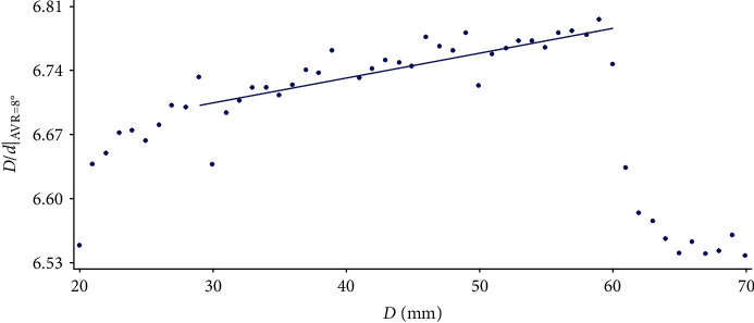
Methods: Data from Raimondi's tables were tabulated on a first step. After this, each column of Raimondi's tables containing values corresponding to vertebral body width (D) were adjusted to a curve determined by AVR = f (d). Third, representative values of each rotation divided by D were obtained through the equation of each column D. In a fourth step, a regression line was fitted to the data in each row, and from its equation, the mean value of the D/d distribution is calculated (value corresponding to the central column, D = 45). Finally, a curve was adjusted to the obtained data using the least squares method. Summary and Conclusion. Our mathematical equation allows the Raimondi method to be used in digital images of any format in a more accurate and simplified approach. This equation can be easily and freely implemented in any CADx system to quantify AVR, providing a more precise use of Raimondi's method, as well as being used in traditional manual measurement as it is performed with Raimondi tables.
Copyright © 2021 José Hurtado-Aviles et al.
Conflict of interest statement
The authors declare that they have no conflicts of interest.
Figures


Similar articles
-
Validity and reliability of a computer-assisted system method to measure axial vertebral rotation.Quant Imaging Med Surg. 2022 Mar;12(3):1706-1715. doi: 10.21037/qims-21-575. Quant Imaging Med Surg. 2022. PMID: 35284293 Free PMC article.
-
Reliability of the axial vertebral rotation measurements of adolescent idiopathic scoliosis using the center of lamina method on ultrasound images: in vitro and in vivo study.Eur Spine J. 2016 Oct;25(10):3265-3273. doi: 10.1007/s00586-016-4492-6. Epub 2016 Mar 7. Eur Spine J. 2016. PMID: 26951170
-
Automatic measurement of vertebral rotation in idiopathic scoliosis.Spine (Phila Pa 1976). 2006 Feb 1;31(3):E80-3. doi: 10.1097/01.brs.0000197653.64796.9d. Spine (Phila Pa 1976). 2006. PMID: 16449892
-
Analysis of four manual and a computerized method for measuring axial vertebral rotation in computed tomography images.Spine (Phila Pa 1976). 2010 May 20;35(12):E535-41. doi: 10.1097/BRS.0b013e3181cb8d2b. Spine (Phila Pa 1976). 2010. PMID: 20489565
-
A review of methods for quantitative evaluation of axial vertebral rotation.Eur Spine J. 2009 Aug;18(8):1079-90. doi: 10.1007/s00586-009-0914-z. Epub 2009 Feb 26. Eur Spine J. 2009. PMID: 19242736 Free PMC article. Review.
Cited by
-
Predicting curve progression for adolescent idiopathic scoliosis using random forest model.PLoS One. 2022 Aug 11;17(8):e0273002. doi: 10.1371/journal.pone.0273002. eCollection 2022. PLoS One. 2022. PMID: 35951527 Free PMC article.
-
Predicting pulmonary function using thoracic deformity parameters in early onset scoliosis patients.PLoS One. 2025 Jul 31;20(7):e0329199. doi: 10.1371/journal.pone.0329199. eCollection 2025. PLoS One. 2025. PMID: 40743255 Free PMC article.
References
-
- Mangone M., Raimondi P., Paoloni M., et al. Vertebral rotation in adolescent idiopathic scoliosis calculated by radiograph and back surface analysis-based methods: correlation between the Raimondi method and rasterstereography. European Spine Journal. 2013;22(2):367–37110. doi: 10.1007/s00586-012-2564-9. - DOI - PMC - PubMed
-
- Nault M.-L., Mac-Thiong J.-M., Roy-Beaudry M., et al. Three-dimensional spinal morphology can differentiate between progressive and nonprogressive patients with adolescent idiopathic scoliosis at the initial presentation. Spine. 2014;39(10):E601–E606. doi: 10.1097/brs.0000000000000284. - DOI - PMC - PubMed
LinkOut - more resources
Full Text Sources
Other Literature Sources
