

Case Reports
doi: 10.1155/2021/6684806.
eCollection 2021.
Gastroduodenal Ulcerative Colitis in Association with Ulcerative Pancolitis
Affiliations
- PMID: 33628535
- PMCID: PMC7895600
- DOI: 10.1155/2021/6684806
Item in Clipboard
Case Reports
Gastroduodenal Ulcerative Colitis in Association with Ulcerative Pancolitis
Case Rep Gastrointest Med.
.
Abstract
Ulcerative colitis (UC) is a chronic inflammatory bowel disease, traditionally regarded as being limited to the colorectum. Although several gastroduodenal lesions have been reported in cases of UC, in general, duodenal lesions in UC are believed to be uncommon and gastric lesions in UC are a rare presentation. In this report, we presented a 66-year-old lady with upper GI presentation with gastroduodenal ulcerative colitis accompanying pancolonic UC.
Copyright © 2021 Khin San Aye et al.
Conflict of interest statement
All the authors have no conflicts of interest to disclose.
Figures
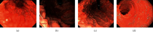
EGD scopy findings; panulcerative gastritis and ulcerative duodenitis. (a) Antrum, (b) fundus, (c) proximal body stomach, and (d) descending part of the duodenum.
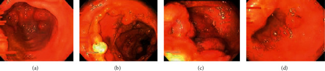
Colonoscopy findings; pancolitis with inflammatory polyps. (a) Rectum, (b) ascending colon, (c) caecum, and (d) terminal ileum.
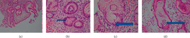
Histologic findings of gastric biopsy; the gastric mucosa shows (a) severe inflammation, ulcerations, and architecture changes, (b) plasmacytosis in the lamina propria, (c) inflammation in the gastric foveolar epithelium, and (d) microabscess formations.
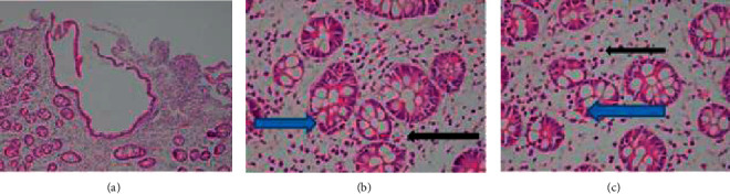
Histologic findings of duodenal biopsy; the duodenum mucosa shows (a) villous atrophy, architecture changes, and edematous lamina propria, (b) plasma cells (black arrow), and (c) cryptitis (blue arrow) in the lamina propria.
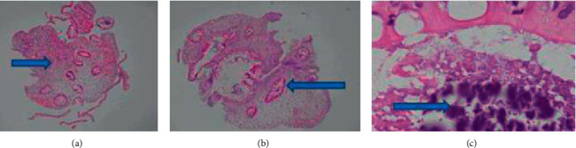
Histologic findings of colon biopsy; all pieces of the colonic polyps show (a) edematous lamina propria, crypt dropout, and mononuclear infiltrates, (b) crypts lined by reactive columnar cells, and (c) bacterial colonies.
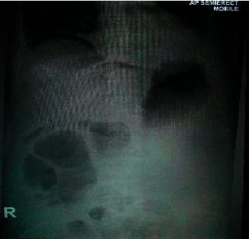
Plain X-ray abdomen; gas under diaphragm positive, gas-filled distended stomach, and some bowel loops.
References
-
- Grossman A. B., Baldassano R. N., Kligman R. M., et al. Chronic Ulcerative Colitis Nelson Textbook of Pediatrics. 19th. Philadelphia, PA, USA: W B Saunders; 2011.
-
- Jang E. S., Lee D. H., Kim J., et al. Age as a clinical predictor of relapse after induction therapy in ulcerative colitis. Hepatogastro-Enterology. 2009;56(94-95):1304–1309. - PubMed
-
- Banerjee R., Pal P., Hilmi I., et al. IBD demographics in Southern Asia. Hyderabad, Telangana: IBD-ENC; 2019.
Publication types
LinkOut - more resources
Full Text Sources
Other Literature Sources
