

IgG4 induces tolerogenic M2-like macrophages and correlates with disease progression in colon cancer
- PMID: 33628623
- PMCID: PMC7889146
- DOI: 10.1080/2162402X.2021.1880687
IgG4 induces tolerogenic M2-like macrophages and correlates with disease progression in colon cancer
Abstract
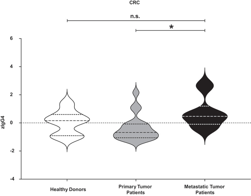
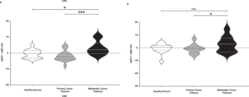
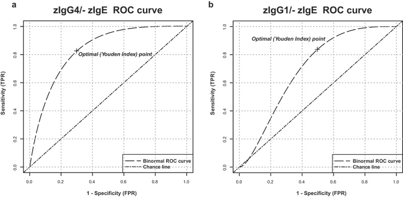
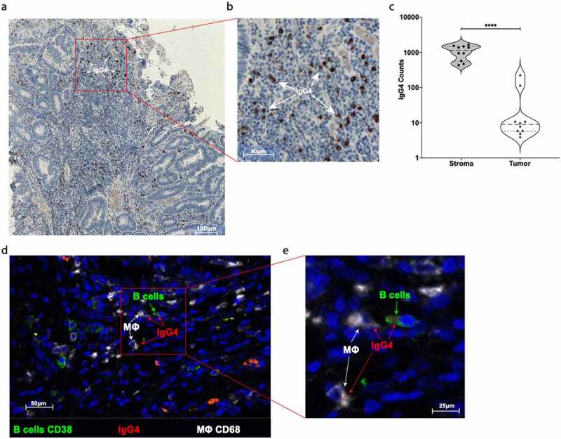
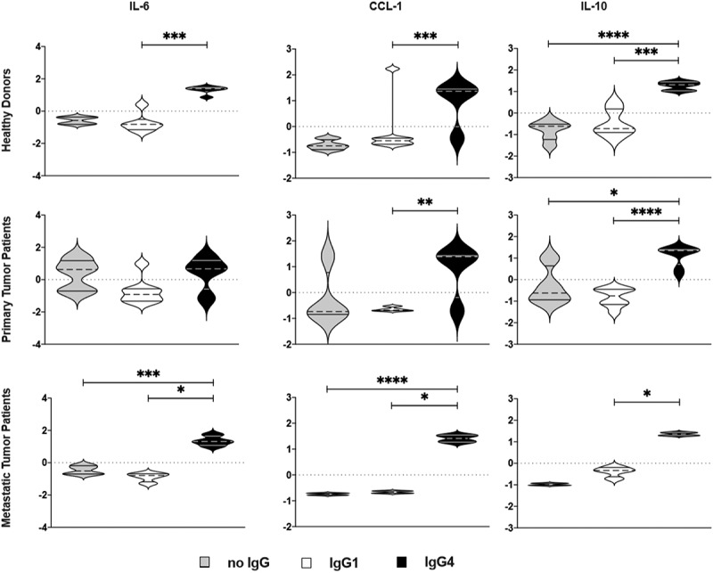
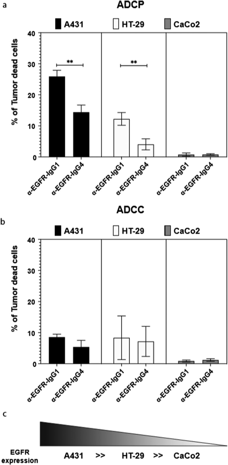
IgG4 subclass antibodies are expressed in alternative Th2 environments featuring high IL-10 expression, including several solid tumors such as melanoma. To induce tolerance, allergen immunotherapy mediates antibody class switching from pro-inflammatory IgE to anti-inflammatory IgG4. We previously reported that IgG4 drives allergic M2 macrophages toward tolerogenic states. Here we assessed the roles of IgG4 and macrophage activation in colorectal cancer (CRC). In this observer-blinded, case-control study, we analyzed total circulating serum IgE, IgG1 and IgG4 levels in CRC (n = 38) patients with (n = 13, TxNxM1) or without (n = 25, TxNxM0) metastasis, and in healthy donors (n = 21). Primary cultures of circulating monocyte-derived macrophages from healthy controls and CRC patients were further evaluated in their responses to stimulation with IgG1 or IgG4. We found higher absolute serum levels of IgG4 in patients with CRC. IgG4 enabled polarization of macrophages derived from CRC patients and healthy controls into alternatively-activated tolerogenic M2b phenotypes. IgG4-stimulated M2 macrophages were characterized by lower surface CD206, CD163, CD14, and CD11b expression and higher CCL-1, IL-10, and IL-6 production. IgG4 was less potent that IgG1 in triggering antibody-dependent cell-mediated phagocytosis (ADCP) of cancer cells. Further, higher z-normalized IgG4/-IgE sera level ratios correlated with the presence of metastasis (p = .0247 and p = .0009, respectively) in CRC patients. High IgG4 in CRC synergizes with macrophages in shaping an immunosuppressive microenvironment and impairs anti-cancer effector cell functions. The shift of serum IgG4/IgE ratios toward enhanced tolerance induction in metastatic disease indicates a role for high IgG4 in disease progression and poor prognostic outcome.
Keywords: IgE; IgG4; M2b; allergooncology; tumor-associated macrophages.
© 2021 The Author(s). Published with license by Taylor & Francis Group, LLC.
Conflict of interest statement
The authors declare no potential conflicts of interest.
Figures
Similar articles
-
IgG4 drives M2a macrophages to a regulatory M2b-like phenotype: potential implication in immune tolerance.Allergy. 2019 Mar;74(3):483-494. doi: 10.1111/all.13635. Epub 2018 Nov 28. Allergy. 2019. PMID: 30338531 Free PMC article.
-
The Role of IgG4 in the Fine Tuning of Tolerance in IgE-Mediated Allergy and Cancer.Int J Mol Sci. 2020 Jul 16;21(14):5017. doi: 10.3390/ijms21145017. Int J Mol Sci. 2020. PMID: 32708690 Free PMC article. Review.
-
Fc-mediated immune stimulating, pro-inflammatory and antitumor effects of anti-HER2 IgE against HER2-expressing and trastuzumab-resistant tumors.J Immunother Cancer. 2025 Mar 12;13(3):e010945. doi: 10.1136/jitc-2024-010945. J Immunother Cancer. 2025. PMID: 40074330 Free PMC article.
-
IgG4-mediated M2 macrophage polarization in tertiary lymphoid structures of esophageal cancer: implications for immunosuppression.Front Immunol. 2025 Jan 17;15:1497783. doi: 10.3389/fimmu.2024.1497783. eCollection 2024. Front Immunol. 2025. PMID: 39896813 Free PMC article.
-
Immunoglobulin G4: an odd antibody.Clin Exp Allergy. 2009 Apr;39(4):469-77. doi: 10.1111/j.1365-2222.2009.03207.x. Epub 2009 Feb 13. Clin Exp Allergy. 2009. PMID: 19222496 Review.
Cited by
-
Systematic exploration of the molecular characteristics of CD8+ T cells to predict the response to immunotherapy and the prognosis of patients with colon adenocarcinoma.Heliyon. 2024 Oct 11;10(23):e39260. doi: 10.1016/j.heliyon.2024.e39260. eCollection 2024 Dec 15. Heliyon. 2024. PMID: 39669138 Free PMC article.
-
Serum IgG4 levels at diagnosis can predict unfavorable outcomes of untreated patients with IgG4-related disease.Sci Rep. 2021 Jun 25;11(1):13341. doi: 10.1038/s41598-021-92814-8. Sci Rep. 2021. PMID: 34172819 Free PMC article.
-
Venom Immunotherapy Does Not Affect Survival of Patients with Malignant Tumor in Poland.J Clin Med. 2024 May 28;13(11):3152. doi: 10.3390/jcm13113152. J Clin Med. 2024. PMID: 38892863 Free PMC article.
-
Tissue macrophages: origin, heterogenity, biological functions, diseases and therapeutic targets.Signal Transduct Target Ther. 2025 Mar 7;10(1):93. doi: 10.1038/s41392-025-02124-y. Signal Transduct Target Ther. 2025. PMID: 40055311 Free PMC article. Review.
-
Case Report: A Programmed Cell Death-1 Inhibitor-Related Abdominal Fibroinflammatory Reaction Affecting Multiple Organs in A Non-Small-Cell Lung Cancer Patient.Front Immunol. 2022 Jul 1;13:874932. doi: 10.3389/fimmu.2022.874932. eCollection 2022. Front Immunol. 2022. PMID: 35860268 Free PMC article.
References
Publication types
MeSH terms
Substances
Grants and funding
LinkOut - more resources
Full Text Sources
Other Literature Sources
Research Materials