

Comparing the Effectiveness of Different Dentinal Desensitizing Agents: In Vitro Study
- PMID: 33628801
- PMCID: PMC7896870
- DOI: 10.1155/2021/6652250
Comparing the Effectiveness of Different Dentinal Desensitizing Agents: In Vitro Study
Abstract
Objectives: To evaluate the in vitro effectiveness of desensitizing agents in reducing dentine permeability.
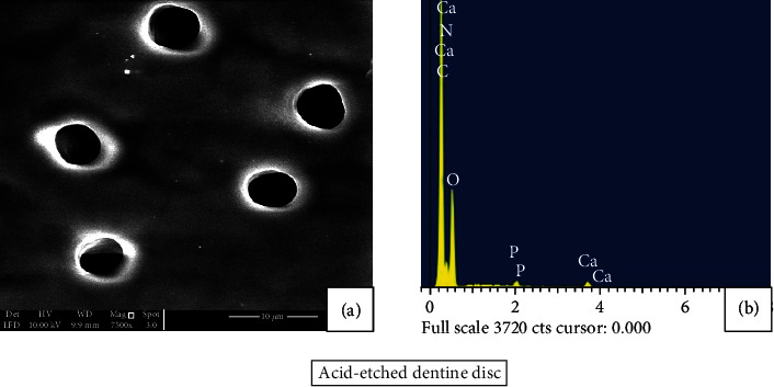
Methods: The efficacy of desensitizing agents in reducing dentine permeability by occluding dentine tubules was evaluated using a fluid filtration device that conducts at 100 cmH2O (1.4 psi) pressure, and SEM/EDX analyses were evaluated and compared. Forty-two dentine discs (n = 42) of 1 ± 0.2 mm width were obtained from caries-free permanent human molars. Thirty dentine discs (n = 30) were randomly divided into 3 groups (n = 10): Group 1: 2.7% wt. monopotassium-monohydrogen oxalate (Mp-Mh oxalate), Group 2: RMGI XT VAR, and Group 3: LIQ SiO2. Dentine permeability was measured following treatment application after 10 minutes, storage in artificial saliva after 10 minutes and 7 days, and citric acid challenge for 3 minutes. Data were analysed with a repeated measures ANOVA test. Dentine discs (n = 12) were used for SEM/EDX analyses to acquire data on morphological changes on dentine surface and its mineral content after different stages of treatment.
Results: Desensitizing agents' application on the demineralized dentine discs exhibited significant reduction of permeability compared to its maximum acid permeability values. Mp-Mh oxalate showed a significant reduction in dentine permeability (p < 0.05) when compared to RMGI XT VAR and LIQ SiO2. On SEM/EDX analysis, all the agents formed mineral precipitates that occluded the dentine tubules.
Conclusions: 2.7% wt. monopotassium-monohydrogen oxalate was significantly effective in reducing dentine permeability compared to RMGI XT VAR and LIQ SiO2.
Copyright © 2021 Mohideen S. Farook et al.
Conflict of interest statement
The authors report no conflicts of interest. The authors alone are responsible for the content and writing of the paper.
Figures


Similar articles
-
Comparison of in vitro dentinal tubule occluding efficacy of two different methods using a nano-scaled bioactive glass-containing desensitising agent.J Dent. 2017 May;60:63-69. doi: 10.1016/j.jdent.2017.03.001. Epub 2017 Mar 4. J Dent. 2017. PMID: 28267581
-
The Effectiveness of Remineralizing Agents on Dentinal Permeability.Biomed Res Int. 2018 Sep 12;2018:4072815. doi: 10.1155/2018/4072815. eCollection 2018. Biomed Res Int. 2018. PMID: 30276206 Free PMC article.
-
Effect of desensitising toothpastes on dentinal tubule occlusion: a dentine permeability measurement and SEM in vitro study.J Dent. 2010 May;38(5):400-10. doi: 10.1016/j.jdent.2010.01.007. Epub 2010 Jan 25. J Dent. 2010. PMID: 20097250
-
The use of calcium-silicate cements to reduce dentine permeability.Arch Oral Biol. 2012 Aug;57(8):1054-61. doi: 10.1016/j.archoralbio.2012.02.024. Epub 2012 Mar 28. Arch Oral Biol. 2012. PMID: 22459650
-
Influence of desensitizing and anti-erosive toothpastes on dentine permeability: An in vitro study.J Dent. 2019 Oct;89:103176. doi: 10.1016/j.jdent.2019.07.014. Epub 2019 Jul 24. J Dent. 2019. PMID: 31351084
References
-
- Addy M. Etiology and clinical implications of dentine hypersensitivity. Dental Clinics of North America. 1990;34(3):503–514. - PubMed
MeSH terms
Substances
LinkOut - more resources
Full Text Sources
Other Literature Sources
