. 2021 Feb 9:2021:6664453.
doi: 10.1155/2021/6664453. eCollection 2021.

Ultrasound May Suppress Tumor Growth, Inhibit Inflammation, and Establish Tolerogenesis by Remodeling Innatome via Pathways of ROS, Immune Checkpoints, Cytokines, and Trained Immunity/Tolerance

Affiliations

Ultrasound May Suppress Tumor Growth, Inhibit Inflammation, and Establish Tolerogenesis by Remodeling Innatome via Pathways of ROS, Immune Checkpoints, Cytokines, and Trained Immunity/Tolerance

Qian Yang et al. J Immunol Res. .

Abstract

Background: The immune mechanisms underlying low-intensity ultrasound- (LIUS-) mediated suppression of inflammation and tumorigenesis remain poorly determined.

Methods: We used microarray datasets from the NCBI GEO DataSet repository and conducted comprehensive data-mining analyses, where we examined the gene expression of 1376 innate immune regulators (innatome genes (IGs) in cells treated with LIUS.

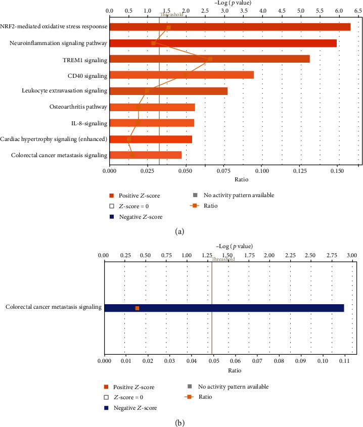
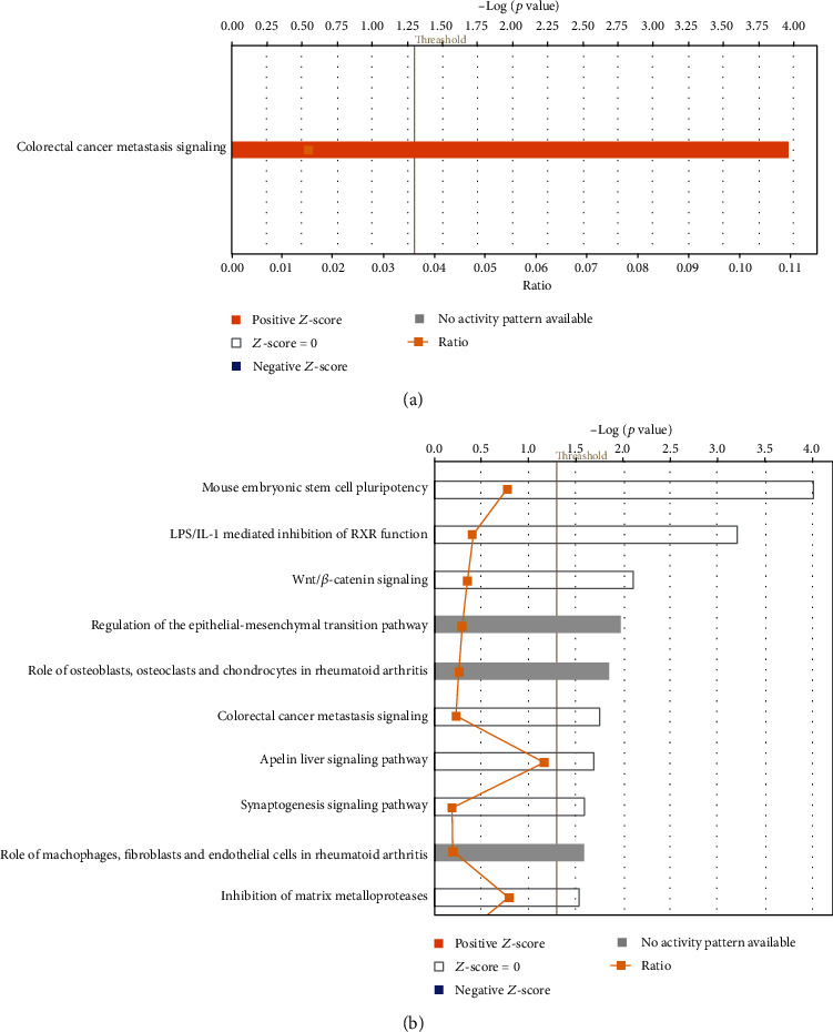
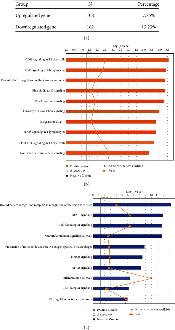
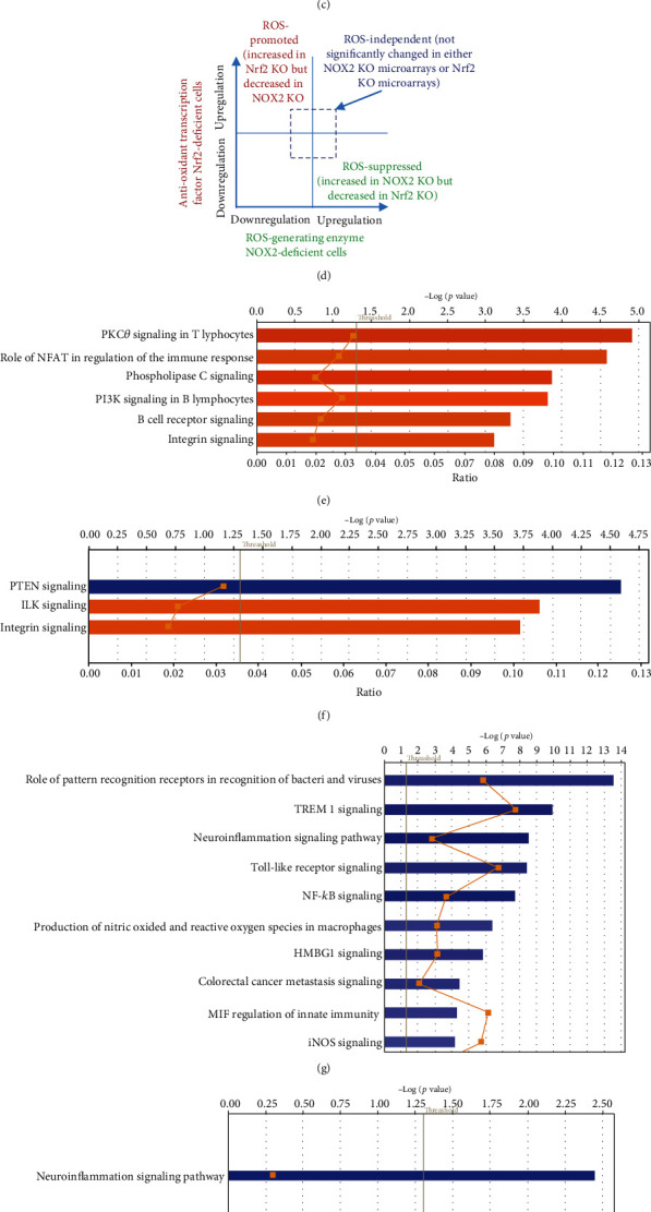
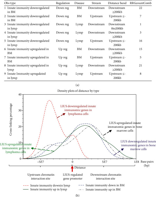
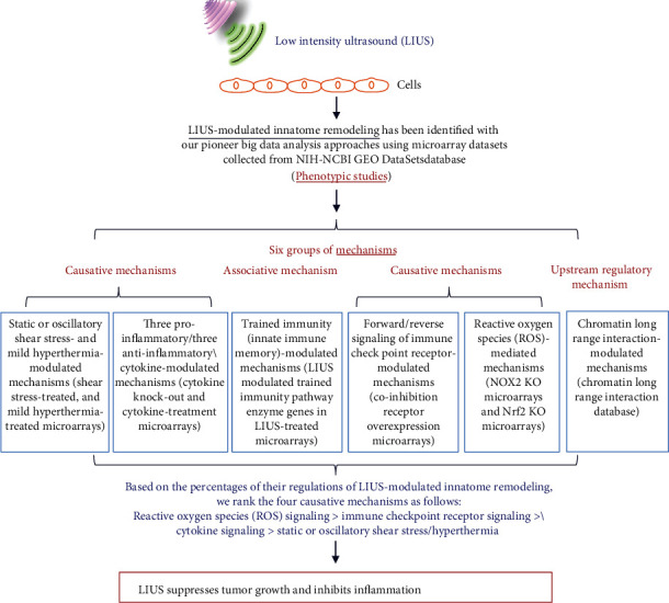
Results: We made the following findings: (1) LIUS upregulates proinflammatory IGs and downregulates metastasis genes in cancer cells, and LIUS upregulates adaptive immunity pathways but inhibits danger-sensing and inflammation pathways and promote tolerogenic differentiation in bone marrow (BM) cells. (2) LIUS upregulates IGs encoded for proteins localized in the cytoplasm, extracellular space, and others, but downregulates IG proteins localized in nuclear and plasma membranes, and LIUS downregulates phosphatases. (3) LIUS-modulated IGs act partially via several important pathways of reactive oxygen species (ROS), reverse signaling of immune checkpoint receptors B7-H4 and BTNL2, inflammatory cytokines, and static or oscillatory shear stress and heat generation, among which ROS is a dominant mechanism. (4) LIUS upregulates trained immunity enzymes in lymphoma cells and downregulates trained immunity enzymes and presumably establishes trained tolerance in BM cells. (5) LIUS modulates chromatin long-range interactions to differentially regulate IGs expression in cancer cells and noncancer cells.

Conclusions: Our analysis suggests novel molecular mechanisms that are utilized by LIUS to induce tumor suppression and inflammation inhibition. Our findings may lead to development of new treatment protocols for cancers and chronic inflammation.


Conflict of interest statement

The authors have no competing interests to disclose.

Figures

Figure 1
Figure 1
(a) Low-intensity ultrasound (LIUS) upregulated 77 out of 1376 (5.6%) innatomic genes and downregulated 39 out of 1376 (2.8%) innatomic genes (IIGs) in human lymphoma U937 cells (GSE10212), suggesting that (1) LIUS increases innatomic gene expressions more than it decreases them in cancer cells in human lymphoma cells, and (2) upregulation of innatomic genes in lymphoma cells serves as a novel immune mechanism underlying antitumor effects of LIUS (see supplemental Table 1 for the detailed gene list. Of note, due to the big data we generated, we have to summarize and present the key findings in this type unconventional format as we previous reported; PMID: 29434588). LIUS upregulated 77 genes that were significantly involved in nine signaling pathways in human lymphoma cells, which included NRF2-mediated oxidative stress response, neuroinflammation signaling, TREM1 signaling, CD40 signaling, leukocyte extravasation signaling, osteoarthritis pathway, IL-8 signaling, cardiac hypertrophy signaling, and cancer metastasis signaling (PMID: 31315034). Of note, eight out of nine pathways were proinflammatory pathways except NRF2 (anti-inflammatory, PMID: 27825853). (b) LIUS downregulated 39 genes that were significantly involved in only one pathway (cancer metastasis signaling) in human lymphoma cells, suggesting that LIUS inhibits inflammation-driven cancer metastasis (PMID: 31315034).
Figure 2
Figure 2
(a) Low-intensity ultrasound (LIUS) upregulates 21 out of 1376 (1.5%) innatomic genes and downregulates 17 out of 1376 (1.2%) innatomic genes in mouse preosteoblast cells (GSE45487), suggesting that LIUS increases innatomic gene expressions slightly more than decreasing them in mouse preosteoblast cells (see supplemental Table 2 for the detailed gene list). LIUS upregulated 21 innatomic genes that were significantly involved in one signaling pathway in mouse preosteoblasts, which is inflammation-driven cancer metastasis signaling. (b) LIUS downregulated 17 innatomic genes that were not significantly involved in any signaling pathway in mouse preosteoblast cells, suggesting that LIUS inhibits innatome in preosteoblasts in multipathways in a nonsignificant manner.
Figure 3
Figure 3
(a) Low-intensity ultrasound (LIUS) upregulated 108 out of 1376 (7.9%) innate immunome genes and downregulated 182 out of 1376 (13.2%) innate immunomic genes in rat bone marrow cells (GSE70662), suggesting that LIUS suppresses innate immunomic gene expressions more than it increases them in bone marrow cells. The LIUS-modulated genes are listed in Supplemental Table 3. (b) LIUS upregulated 108 genes that were significantly involved in 54 signaling pathways in bone marrow cells. The top ten pathways include CD28 signaling in T cells, PI3K signaling in B lymphocytes, the role of NFAT in regulation of immune responses, phospholipase C signaling, B cell receptor signaling, leukocyte extravasation signaling, integrin signaling, PKC zeta signaling in T lymphocytes, ICOS-ICOSL signaling in T helper cells, and non-small-cell lung cancer signaling. (c) LIUS downregulated 182 genes that were significantly involved in 70 signaling pathways in bone marrow cells. The top ten pathways include the role of pattern recognition receptors, TREM1 signaling, Toll-like receptor signaling, neuroinflammation signaling, production of nitric oxide and reactive oxygen species (ROS) in macrophages, HMGB1 signaling, NF-κB signaling, inflammation pathway, B cell receptor signaling, and MIF regulation of innate immunity. These results suggest that LIUS inhibits numerous key innate immunity and inflammation pathways in bone marrow cells, which may be responsible for LIUS therapeutic effects of inflammation suppression since nine out of the top ten pathways are related to danger signal recognition and inflammation initiation.
Figure 4
Figure 4
(a) The Venn Diagram analyses shows that LIUS-upregulated innatomic genes in three cell types are partially shared. The three genes shared by lymphoma cells (L) and preosteoblasts (P) and the 11 genes shared by lymphoma cells (L) and bone marrow cells (B) may be used for LIUS therapeutic markers. However, the majority of LIUS-upregulated innatomic genes are cell type specific. (b) The Venn Diagram analyses shows that the signaling pathways involved in LIUS-upregulated innatomic genes in three cell types are partially shared, such as metastasis signaling. In addition, three pathways are shared by LIUS-treated lymphoma cells and bone marrow cells including leukocyte extravasation, osteoarthritis pathway, and cardiac hypertrophy signaling. However, the majority of LIUS-upregulated innatomic pathways in lymphoma and bone marrow cells are cell type specific. (c) The Venn Diagram analyses shows that LIUS-downregulated innatomic genes in three cell types are not shared. The majority of LIUS-downregulated innatomic genes are cell type specific (L: lymphoma cells; B: bone marrow cells; P: preosteoblasts). (d) The Venn Diagram analysis shows that the signaling pathways involved in LIUS-downregulated innatomic genes in lymphoma cells and bone marrow cells are partially shared, such as metastasis signaling. However, the majority of LIUS-downregulated innatomic pathways in bone marrow cells are cell type specific.
Figure 5
Figure 5
Figure 6
Figure 6
Figure 7
Figure 7
(a) LIUS upregulated trained immunity enzymes in lymphoma cells and downregulated trained immunity enzymes in bone marrow cells, suggesting that LIUS enhances innate immune responses in cancer cells and inhibits innate immune responses in noncancer cells, which are associated with its modulation of trained immunity enzyme expressions. By analyzing microarray data of LIUS-treated human lymphoma cells, mouse preosteoblasts, and rat bone marrow cells, we examined a novel hypothesis that LIUS induces its therapeutic effects in cells by modulating the expressions of our newly reported (PMID: 31153039) innate immune memory (trained immunity) pathway enzymes. Our results show that LIUS downregulates 12 out of 102 trained immunity genes, including 11 (10.8%) in bone marrow cells and one in lymphoma cells. In addition, LIUS induces 11 out of 102 trained immunity genes (10.8%) including 6 in lymphoma cells, 1 in preosteoblasts, and 4 in bone marrow cells. Moreover, in bone marrow cells, LIUS upregulates 4 trained immunity genes and downregulates 11 trained immunity genes including 8 glycolysis enzymes, one acetyl-CoA enzyme, and two mevalonate pathway enzymes, suggesting that 2.75-fold more downregulation than upregulation of trained immunity genes in bone marrow cells contributes significantly to LIUS-suppressed innate immunity and inflammation. Finally, in contrast, in lymphoma cells, LIUS upregulation of six trained immunity enzymes including 5 glycolysis enzymes and one acetyl-CoA generation enzymes and downregulation of one glycolysis enzyme in lymphoma cells (6-fold more upregulation than downregulation) contribute significantly to LIUS-induced innate immunity enhancement. (b) Low-intensity ultrasound (LIUS) therapy upregulated trained immunity enzymes (PMID: 27102489) in lymphoma cells and downregulated trained immunity enzymes in bone marrow cells. These results suggest that LIUS enhances antitumor immune responses against lymphoma cells at least partially by upregulating trained immunity pathways, which is similar to that proposed by others (PMID: 27903713); LIUS also inhibits inflammation in noncancer cells by suppressing trained immunity pathways (see our recent report, PMID: 31153039). Trained immunity (innate immune memory): newly characterized adaptive metabolic and epigenetic remodeling of innate immune cells including (1) upregulation of glycolysis pathway enzymes, (2) increased acetyl-CoA generation, (3) upregulation of mevalonate pathway enzymes, and (4) remodeling of histone methylation and acetylation.
Figure 8
Figure 8
Figure 9
Figure 9
(a) LIUS modulates oxidative stress-related gene expressions (reactive oxygen species, ROS, and regulatome). Three out of a total of 84 (3.70%) oxidative stress genes (ROS regulatome) were upregulated in lymphoma cells (L) and bone marrow cells (B). Gpx3 was shared in L and B. Two out of a total of 84 genes (2.47%) were downregulated in bone marrow cells (B). No oxidative stress gene expressions were changed in the preosteoblast microarray dataset. (b) LIUS modulates the ROS regulatome in a cell-specific manner. (c) The Ingenuity Pathway Analyses (IPA) of ROS dependence to LIUS-modulated innatomic genes in three types of cells were summarized into four groups: (1) the ROS-promoted group, (2) the ROS-suppressed group, (3) the ROS-dependent/suppressed pathway-uncertain group, and (4) the ROS-independent group. These results demonstrated that LIUS-modulated innatomic genes were significantly mediated by ROS-promoted or ROS-suppressed pathways in bone marrow cells, which were higher than those of LIUS-modulated innatomic genes in lymphoma cells. In addition, the IPA-identified significant pathway numbers in bone marrow cells that were significantly higher than those of lymphoma cells, suggesting that ROS-modulated genes in lymphoma cells are much more diversified in the pathways than those of bone marrow cells (see supplemental Tables 17A-17F for details). (d) LIUS-modulated innatomic genes were classified into four groups, namely, (1) the ROS-promoted group, (2) the ROS-suppressed group, (3) the uncertain (not shown) group, and (4) the ROS-independent group, based on their expression changes in ROS-generating enzyme NOX2 KO microarrays and antioxidant transcription factor Nrf2 KO microarrays. (e) Six ROS-promoted pathways were involved in LIUS-upregulated genes in bone marrow cells. (f) Three ROS-suppressed pathways were involved in LIUS-upregulated genes in bone marrow cells. (g) The top 10 of a total of 31 ROS-promoted pathways were involved in LIUS-downregulated genes in bone marrow cells. (h) Only one ROS-suppressed pathway was involved in LIUS-downregulated genes in bone marrow cells. (I) Only one ROS-suppressed pathway was involved in LIUS-upregulated genes in lymphoma cells. (j) LIUS modulates innatome via reactive oxygen species (ROS) pathways mediated by pro-ROS generation enzyme nicotinamide adenine dinucleotide phosphate (NADPH) oxidase 2 (NOX2) (PMIDs: 21629295; 28916473) and antioxidant transcription factor nuclear erythroid-2 like factor (Nrf2) (PMID: 30610225).
Figure 10
Figure 10
(a) Innate immunome chromatin looping makes long-range interactions that can be regulated by low-intensity ultrasound (LIUS). (a) Chromatin is a whole structure of complex DNA and proteins; it forms the chromosomes of eukaryotic organisms and is packaged inside the nucleus. Nucleosome is a basic unit of chromatin, consisting of a length of DNA coiled around a core of histones. (B) Chromatin looping makes gene promoter and distal regulatory elements come in close proximity and possibly interact with each other, which can be regulated by LIUS. (c) Long-range interactions allow communication between promoters and different distant regulatory elements (for a better understanding, please refer to Figure 4 of our recent paper published on Frontiers in Oncology 2019 at https://www.frontiersin.org/articles/10.3389/fonc.2019.00600/full). (b) LIUS modulates chromatin long-range interactions to regulate innatomic gene expressions in lymphoma cells (cancer cells) and bone marrow cells (the numbers of LIUS-regulated innatomic genes in preosteoblast cells were low so that chromatin long-range interaction data were too low to be analyzed). These results show that (i) the chromosome interaction zones are mostly located downstream of LIUS-upregulated innatomic genes in lymphoma cells, but the chromosome interaction zones are located in similar numbers both upstream and downstream of LIUS-upregulated genes in noncarcinoma cells, and (ii) the long-range interaction zones of LIUS-upregulated genes in lymphoma cells are located in a more concentrated manner both upstream and downstream (between 102 base pairs (bp) and 108 bp) than those of noncancer cells.
Figure 11
Figure 11
A new working model on LIUS-mediated cancer suppression and anti-inflammatory mechanisms via modulating innatome.

Similar articles

Cited by

References

    1. de Castro C. C., Gondim Gomes C. B., Martins M. R., de Sousa J. C., Magalhaes S. M., Pinheiro R. F. Tissue Doppler echocardiography detects preclinical markers of cardiac lesion in MDS patients. Journal of Hematology & Oncology. 2012;5(1):p. 30. doi: 10.1186/1756-8722-5-30. - DOI - PMC - PubMed
    1. Yang Q., Nanayakkara G. K., Drummer C., et al. Low-intensity ultrasound-induced anti-inflammatory effects are mediated by several new mechanisms including gene induction, immunosuppressor cell promotion, and enhancement of exosome biogenesis and docking. Frontiers in Physiology. 2017;8:p. 818. doi: 10.3389/fphys.2017.00818. - DOI - PMC - PubMed
    1. Schroeder A., Kost J., Barenholz Y. Ultrasound, liposomes, and drug delivery: principles for using ultrasound to control the release of drugs from liposomes. Chemistry and Physics of Lipids. 2009;162(1-2):1–16. doi: 10.1016/j.chemphyslip.2009.08.003. - DOI - PubMed
    1. Sheikov N., McDannold N., Vykhodtseva N., Jolesz F., Hynynen K. Cellular mechanisms of the blood-brain barrier opening induced by ultrasound in presence of microbubbles. Ultrasound in Medicine & Biology. 2004;30(7):979–989. doi: 10.1016/j.ultrasmedbio.2004.04.010. - DOI - PubMed
    1. Forbes M. M., O’Brien W. D., Jr. Development of a theoretical model describing sonoporation activity of cells exposed to ultrasound in the presence of contrast agents. The Journal of the Acoustical Society of America. 2012;131(4):2723–2729. doi: 10.1121/1.3687535. - DOI - PMC - PubMed

MeSH terms