. 2021 Mar;9(1):23-33.
Epub 2021 Jan 25.

Differential Expression Profiles of Mitogenome Associated MicroRNAs Among Colorectal Adenomatous Polyps

Affiliations

Differential Expression Profiles of Mitogenome Associated MicroRNAs Among Colorectal Adenomatous Polyps

LaShanale Wallace et al. Cancer Res J (N Y N Y). 2021 Mar.

Abstract

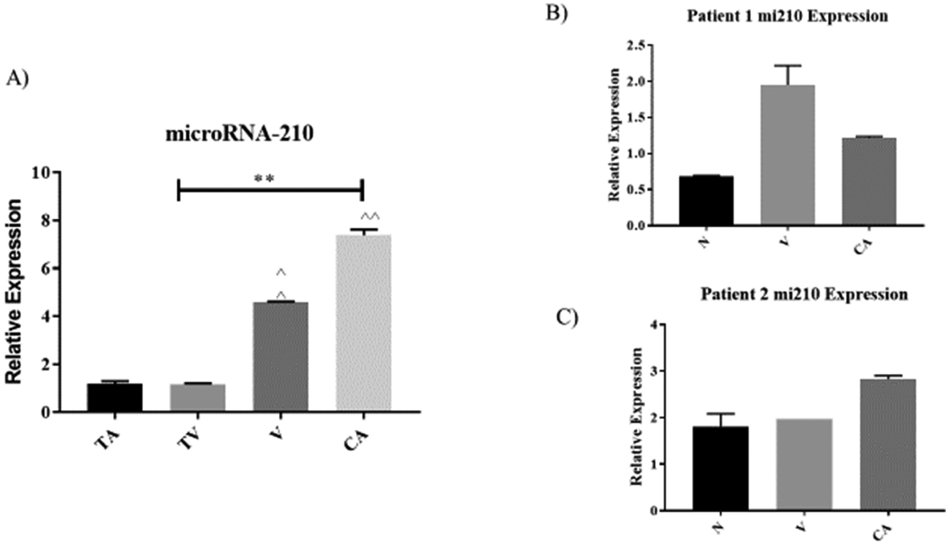
Colorectal tumors are mostly of epithelial origin and represent a wide spectrum of neoplasms. About 97% of colorectal cancer originating from benign lesions of adenomatous polyps are adenocarcinomas. Reactive oxygen species (ROS) generating from mitochondrial DNA (mtDNA) mutations and microRNAs (miRNAs) are associated with oncogene and tumor suppressor genes regulation which are known to parallel the tissue abnormalities involved with tumorigenesis such as colorectal adenoma to adenocarcinoma. However, the differential expression patterns of mitochondrial associated microRNAs (referred as MitomiRs) among colorectal adenomatous polyps progression is yet to be determined. Thus, the aim of this study was to determine the differential expressions profiles of MitomiRs (miR-24, miR-181, miR-210, miR-21 and miR378) in patients with colorectal adenomatous polyps tissues in correlation with clinicopathological tumor architectures of tubular, tubulovillous, villous adenomas and adenocarcinomas. Isolation of mitochondria RNA from colorectal adenomatous polyps, adenocarcinomas, and normal adjacent tissue samples was performed and assessed for mitochondrial associated miRNAs expression differences using quantitative reverse transcription PCR. Data from this study demonstrates that mitochondria genome expression of mitomiRNAs; miR-24, miR-181, miR-210, miR-21 and miR-378 in colorectal tissue samples varies among the adenomatous polyps. Expression of mitomiRNAs 24, 181, 210 and 378 progressively increased from the precancerous of adenomatous polyps to adenocarcinoma. In addition, miR-210 and miR-181 expression increased 3 folds in villous adenomas and greater than 3 folds increased in miR378 in adenocarcinoma (p < 0.005) when compared to tubular adenoma. Meanwhile, miR-21 increased progressively in adenoma tissues but decreased almost 2.5 folds in adenocarcinomas when compared to villous adenoma tissues (p < 0.001). These results suggest mitomiRs may regulate important mitochondrial functional pathways leading to a more favorable environment for transformation or progression of colorectal adenomatous polyps into adenocarcinomas.

Keywords: CRC Tissues; Colorectal Adenomas; Mitochondrial microRNA (Denoted: mitomiRNAs); Reactive Oxygen Species.


Conflict of interest statement

Conflicts of Interest The authors declare no conflict of interest.

Figures

Figure 1.
Figure 1.
The relative expression levels of MitomiRNA-24 observed among patient tissue samples (n=64 pairs) of colorectal adenomas (tubular (T), tubulovillous (TV), villous (V)), pooled adenocarcinoma (CA) and their normal adjacent tissues (N). A) Correlation of each clinicopathological features of colorectal adenomas and pooled adenocarcinoma tissues with expression levels of mitomiRNA-24 in extracted mitochondria RNA. Mean expression values from each tissue histological type were pooled and normalized to their paired normal adjacent tissues. Values are presented as relative expressions of mitomiRNA-24 which showed markedly increased progressively from colorectal adenomas to the pooled adenocarcinomas. B) Comparison of mitomiRNA-24 expression patterns in normal adjacent, villous, and adenoma tissues based on individual patient. Patient one is defined as 77 years old white female with stage I colon cancer. C) Comparison of mitomiRNA-24 expression patterns in 89 years old white female with stage II colon cancer. Bar diagrams show mean ± from pooled results. Asterisk (٭) P<0.005.
Figure 2.
Figure 2.
The relative expression levels of MitomiRNA-181 observed among patient tissue samples (n=64 pairs) of colorectal adenomas (tubular (T), tubulovillous (TV), villous (V)), pooled adenocarcinoma (CA) and normal adjacent tissues (N). A) Correlation of each clinicopathological features of colorectal adenomas and adenocarcinoma tissues with expression levels of mitomiRNA-181 in extracted mitochondria RNA. Mean expression values from each tissue histological type were pooled and normalized to their paired normal adjacent tissues. Values are presented as relative expressions of mitomiRNA-181 which showed markedly increased progressively from colorectal adenomas to the pooled adenocarcinomas. B) Comparison of mitomiRNA-181 expression patterns in normal, villous, and adenoma tissues based on individual patient. Patient one is defined as 77 years old white female with stage I colon cancer. C) Comparison of mitomiRNA-181 expression patterns in 89 years old white female with stage II colon cancer. Bar diagrams show mean ± from pooled results. Asterisk (٭) P<0.005.
Figure 3.
Figure 3.
The relative expression levels of MitomiRNA-21 observed among patient tissue samples (n=64 pairs) of colorectal adenomas: (tubular (T), tubulovillous (TV), villous (V)), pooled adenocarcinoma (CA) and normal adjacent tissues (N). A) Correlation of each clinicopathological features of colorectal adenomas and adenocarcinoma tissues with expression levels of mitomiRNA-21 in extracted mitochondria RNA. Mean expression values from each tissue histological type were pooled and normalized to their paired normal adjacent tissues. Values are presented as relative expressions of mitomiRNA-21 which showed markedly increased colorectal villous adenomas only compared to tubular, tubulovillous and pooled adenocarcinomas. B) Comparison of mitomiRNA-21 expression patterns in normal, villous and adenoma tissues based on individual patient. Patient one is defined as 77 years old white female with stage I colon cancer. C) Comparison of mitomiR expression patterns in 89 years old white female with stage II colon cancer. Bar diagrams show mean ± from pooled results. Asterisk (٭٭) P<0.001.
Figure 4.
Figure 4.
The relative expression levels of MitomiRNA-210 observed among patient tissue samples (n=64 pairs) of colorectal adenomas, (tubular (T), tubulovillous (TV), villous (V)), to the pooled adenocarcinoma (CA) and normal surrounding tissues (N). A) Correlation of each clinicopathological features of colorectal adenomas and adenocarcinoma tissues with expression levels of mitomiRNA-210 in extracted mitochondria RNA. Mean expression values form each tissue histological type were pooled and normalized to their paired normal adjacent tissues. Values are presented as relative expressions of mitomiRNA-210 which showed markedly increased progressively from colorectal adenomas to adenocarcinomas. B) Comparison of mitomiRNA-210 expression patterns in normal, villous, and adenoma tissues based on individual patient. Patient one is defined as 77 years old white female with stage I colon cancer. C) Comparison of mitomiRNA-210 expression patterns in 89 years old white female with stage II colon cancer. Bar diagrams show mean ± from pooled results. Asterisk (٭٭) P<0.001.
Figure 5.
Figure 5.
The relative expression levels of MitomiRNA-378 observed among patient tissue samples (n=64 pairs) of colorectal adenomas. (tubular (T), tubulovillous (TV), villous (V)), to the pooled adenocarcinoma (CA) and normal adjacent tissues (N). A) Correlation of each clinicopathological features of colorectal adenomas and adenocarcinoma tissues with expression levels of mitomiRNA-378 in extracted mitochondria RNA. Mean expression values from each tissue histological type were pooled and normalized to their paired normal adjacent tissue. Values are presented as relative expressions of mitomiRNA-378 which showed markedly increased progressively from the colorectal adenomas to the pooled adenocarcinomas. B) Comparison of mitomiRNA-378 expression patterns in normal, villous, and adenoma tissues based on individual patient. Patient one is defined as 77 years old white female with stage I colon cancer. C) Comparison of mitomiRNA-378 expression patterns in 89 years old white female with stage II colon cancer.
Figure 6.
Figure 6.
Schematic of mitomiRNAs interactions with mitochondrial encoded genes either positive or negative suggestively. Image was created using the Pathway Studio program and arrows indicate flow through signaling pathways; touching boxes indicate direct binding of mitomiRNAs signaling components. Data from various articles has shown that microRNAs have both positive and negative roles on subunits involved in oxidative phosphorylation. Within this interaction, reactive oxygen species may also play a role on the ability for mentioned microRNAs classification as an oncomiR or tumor suppressor.

References

    1. Rawla P; Sunkara T; Barsouk A Epidemiology of colorectal cancer: incidence, mortality, survival, and risk factors. Prz Gastroenterol. 2019, 14 (2): 89–103. - PMC - PubMed
    1. Bujanda L; Cosme A; Gil I; Juan I; Arenas-Mirave JI, Malignant colorectal polyps. World J Gastroenterol. 2010; 16, (25). - PMC - PubMed
    1. Munteanu I; Mastalier B Genetics of colorectal cancer. J Med Life. 2014; 7 (4), 507–511. - PMC - PubMed
    1. Sun D; Yu F; Ma Y; Zhao R; Chen X; Zhu J; Zhang CY.; Chen JZ,; Hang J MicroRNA-31 activates the RAS pathway and functions as an oncogenic MicroRNA in human colorectal cancer by repressing RAS p21 GTPase activating protein 1 (RASA1). The Journal of biological chemistry. 2013, 288 (13), 9508–9518. - PMC - PubMed
    1. Kent O; Mendell J; Rottapel R Transcriptional Regulation of miR-31 by Oncogenic KRAS Mediates Metastatic Phenotypes by Repressing RASA1. Molecular Cancer Research: MCR 2016, 14 (3), 267–277. - PMC - PubMed

LinkOut - more resources