

Case Reports
doi: 10.1080/23320885.2021.1884560.
Intravascular papillary endothelial hyperplasia of the finger: a case of Masson's tumor
Affiliations
- PMID: 33628865
- PMCID: PMC7889222
- DOI: 10.1080/23320885.2021.1884560
Item in Clipboard
Case Reports
Intravascular papillary endothelial hyperplasia of the finger: a case of Masson's tumor
Case Reports Plast Surg Hand Surg.
.
Abstract
Intravascular papillary endothelial hyperplasia is an uncommon benign vascular lesion characterized by a reactive proliferation of endothelial cells. The lesion of the finger often presents diagnostic challenges to surgeons because of its rarity. We report a case of intravascular papillary endothelial hyperplasia to facilitate the recognition of this uncommon lesion.
Keywords: Blood vessels; cysts; endothelium; fingers; hyperplasia.
© 2021 The Author(s). Published by Informa UK Limited, trading as Taylor & Francis Group.
Conflict of interest statement
No potential conflict of interest was reported by the author(s).
Figures
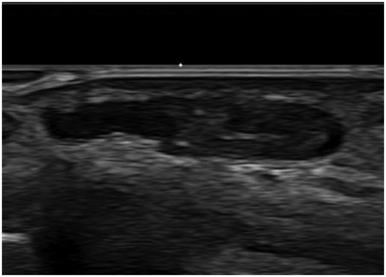
Preoperative ultrasonography reveals about 0.7 × 0.3 × 1.2 cm sized cystic lesion with no remarkable increased vascularity.

Intraoperative photograph shows a soft, red purple, and well-circumscribed cystic mass looking like a hemangioma at the volar surface of left 2nd proximal phalanx.

Histopathologic appearance of the lesion. (a) The specimen showed intravascular proliferative lesion with organizing thrombus (arrow) (H&E, ×15). (b) High magnification of papillary proliferations revealed fibrinous and hyaline stalks lined by endothelial cells (H&E, ×400). (c) These lining cells were positive for anti-CD31 (immunohistochemical stain, clone JC70, Cell Marque, ×100).
Similar articles
-
Intravascular papillary endothelial hyperplasia (Masson's tumor) of the finger: a case report and review of the literature.Case Reports Plast Surg Hand Surg. 2025 Jun 5;12(1):2513066. doi: 10.1080/23320885.2025.2513066. eCollection 2025. Case Reports Plast Surg Hand Surg. 2025. PMID: 40487413 Free PMC article.
-
Intravascular Papillary Endothelial Hyperplasia: Case Report of a Recurrent Masson's Tumor of the Finger and Review of Literature.J Hand Microsurg. 2021 Jul;13(3):164-168. doi: 10.1055/s-0039-3401381. Epub 2020 Jan 16. J Hand Microsurg. 2021. PMID: 34602798 Free PMC article.
-
An intravascular papillary endothelial hyperplasia of the hand radiologically mimicking a hemangiopericytoma: A case report and literature review.SAGE Open Med Case Rep. 2018 Jan 10;6:2050313X17752851. doi: 10.1177/2050313X17752851. eCollection 2018. SAGE Open Med Case Rep. 2018. PMID: 29348916 Free PMC article.
-
Masson's tumor of the breast: Rare differential for new or recurrent breast cancer-Case report, pathology, and review of the literature.Breast J. 2020 Apr;26(4):752-754. doi: 10.1111/tbj.13608. Epub 2019 Sep 20. Breast J. 2020. PMID: 31538368 Review.
-
Intravascular papillary endothelial hyperplasia in the oral mucosa and jawbones: A collaborative study of 20 cases and a systematic review.J Oral Pathol Med. 2021 Jan;50(1):103-113. doi: 10.1111/jop.13127. Epub 2020 Dec 7. J Oral Pathol Med. 2021. PMID: 33188642
Cited by
-
Intravascular Papillary Endothelial Hyperplasia (Masson Tumor) of the Right Thumb: A Case Report and Literature Review.Plast Reconstr Surg Glob Open. 2023 Sep 7;11(9):e5224. doi: 10.1097/GOX.0000000000005224. eCollection 2023 Sep. Plast Reconstr Surg Glob Open. 2023. PMID: 37691699 Free PMC article.
-
Intravascular papillary endothelial hyperplasia (masson tumor) of the right wrist: A case report and literature review.JPRAS Open. 2024 Apr 6;41:240-243. doi: 10.1016/j.jpra.2024.03.014. eCollection 2024 Sep. JPRAS Open. 2024. PMID: 39113727 Free PMC article.
-
Intravascular papillary endothelial hyperplasia (Masson's tumor) of the finger: a case report and review of the literature.Case Reports Plast Surg Hand Surg. 2025 Jun 5;12(1):2513066. doi: 10.1080/23320885.2025.2513066. eCollection 2025. Case Reports Plast Surg Hand Surg. 2025. PMID: 40487413 Free PMC article.
-
Masson's Hemangioma of Knee: A Rare Case Report.J Orthop Case Rep. 2021 Aug;11(8):79-83. doi: 10.13107/jocr.2021.v11.i08.2376. J Orthop Case Rep. 2021. PMID: 35004382 Free PMC article.
References
-
- Masson P. Hemangioendothelioma vegetant intravasculaire. Bull Soc Anat. 1923;93:517–523.
-
- Henschen F. L'endovasculite proliferante thrombopoietique dans la lesion vasculaire locale. Ann Anat Patho. 1932;9:113–121.
-
- Clearkin K, Enzinger F.. Intravascular papillary endothelial hyperplasia. Arch Pathol Lab Med. 1976;100:441–444. - PubMed
-
- Tedla M, Bežová M, Biró C, et al. . Intravascular papillary endothelial hyperplasia of larynx: case report and literature review of all head and neck cases. Otolaryngol Pol. 2014;68:200–203. - PubMed
-
- Requena L, Sangueza OP.. Cutaneous vascular proliferation. Part II. Hyperplasias and benign neoplasms. J Am Acad Dermatol. 1997;37:887–919. - PubMed
Publication types
LinkOut - more resources
Full Text Sources
Other Literature Sources