

A case study of methadone-induced delayed post-hypoxic leukoencephalopathy with improvement by antioxidant therapy
- PMID: 33629035
- PMCID: PMC7881437
- DOI: 10.14744/nci.2020.29795
A case study of methadone-induced delayed post-hypoxic leukoencephalopathy with improvement by antioxidant therapy
Abstract
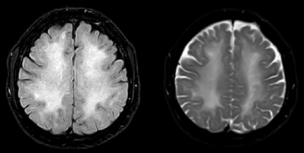
Delayed post-hypoxic leukoencephalopathy (DPHL) is a syndrome that may occur as a result of the hypoxic event, including opiate overdose. The pathophysiology of this entity is not fully known. Within a neuropsychiatric context, the diagnosis of this rare disease is important. A 39-year-old man with a history of methadone overdose presented with loss of consciousness and fever. After clinical evaluations, laboratory analysis, including various tests on blood and cerebrospinal fluid and magnetic resonance imaging, the patient was diagnosed with methadone-induced DPHL. Treatment with antioxidants, including vitamins E, C and B complex, produced a favorable outcome. In rare cases, methadone overdose may lead to DPHL. Antioxidants therapy should be considered in the treatment of this rare disorder.
Keywords: Antioxidants; anoxia; delayed post-hypoxic leukoencephalopathy; hypoxia; leukoencephalopathy; methadone; overdose; vitamin.
Copyright: © 2021 by Istanbul Northern Anatolian Association of Public Hospitals.
Conflict of interest statement
Conflict of Interest: No conflict of interest was declared by the authors.
Figures

Similar articles
-
Delayed Posthypoxic Leukoencephalopathy: Improvement with Antioxidant Therapy.Case Rep Neurol. 2015 Dec 24;7(3):242-6. doi: 10.1159/000441892. eCollection 2015 Sep-Dec. Case Rep Neurol. 2015. PMID: 26955335 Free PMC article.
-
Positive 14-3-3 protein in cerebrospinal fluid followed by poppy-induced delayed post-hypoxic leukoencephalopathy: A case report.Heliyon. 2024 Aug 30;10(17):e37129. doi: 10.1016/j.heliyon.2024.e37129. eCollection 2024 Sep 15. Heliyon. 2024. PMID: 39296161 Free PMC article.
-
A case report of long-term effects of Delayed post-hypoxic leukoencephalopathy (DPHL) following benzodiazepine overdose.Clin Neuropsychol. 2024 Oct;38(7):1756-1772. doi: 10.1080/13854046.2024.2315746. Epub 2024 Feb 20. Clin Neuropsychol. 2024. PMID: 38378478
-
Acute fatal posthypoxic leukoencephalopathy following benzodiazepine overdose: a case report and review of the literature.BMC Neurol. 2015 Apr 30;15:69. doi: 10.1186/s12883-015-0320-6. BMC Neurol. 2015. PMID: 25925073 Free PMC article. Review.
-
Delayed posthypoxic leukoencephalopathy: a case series and review of the literature.Brain Behav. 2015 Aug;5(8):e00364. doi: 10.1002/brb3.364. Epub 2015 Jul 3. Brain Behav. 2015. PMID: 26357591 Free PMC article. Review.
Cited by
-
Spectrum of delayed post-hypoxic leukoencephalopathy syndrome: A systematic review.World J Clin Cases. 2024 Oct 16;12(29):6285-6301. doi: 10.12998/wjcc.v12.i29.6285. World J Clin Cases. 2024. PMID: 39417054 Free PMC article.
References
Publication types
LinkOut - more resources
Full Text Sources