

Ultrasound-Guided Minimal Invasive Carpal Tunnel Release: An Optimized Algorithm
- PMID: 33629135
- PMCID: PMC8172390
- DOI: 10.1007/s00270-021-02789-2
Ultrasound-Guided Minimal Invasive Carpal Tunnel Release: An Optimized Algorithm
Abstract
Purpose: To present a safety-optimized ultrasound-guided minimal invasive carpal tunnel release (CTR) procedure.
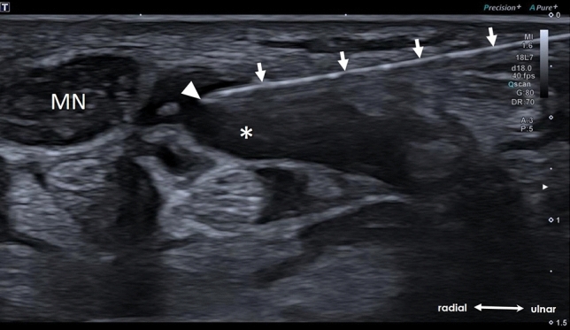
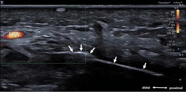
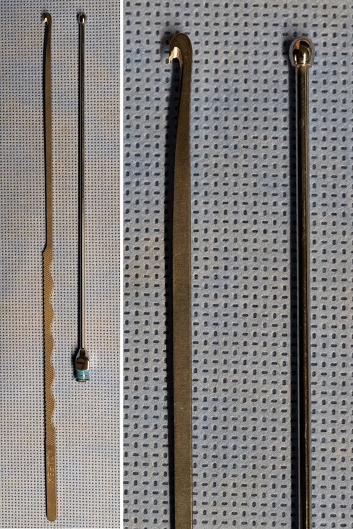
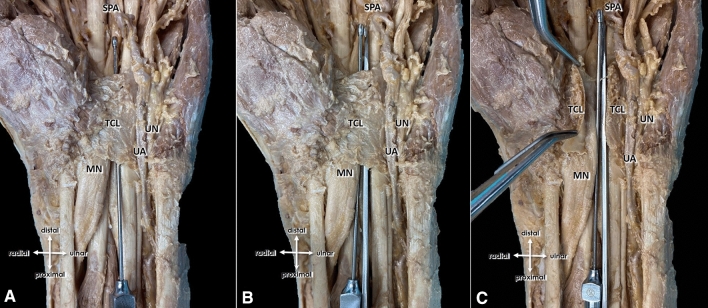
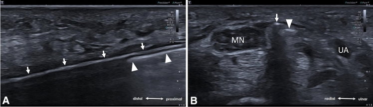
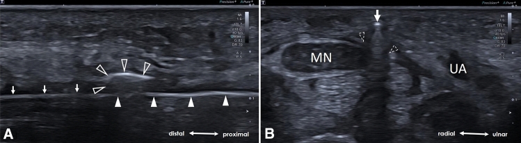
Materials and methods: 104 patients (67 female, 37 male; mean age 60.6 ± 14.3 years, 95% CI 57.9 to 63.4 years) with clinical and electrophysiological verified typical carpal tunnel syndrome were referred for a high-resolution ultrasound of the median nerve and were then consecutively assigned for an ultrasound-guided CTR after exclusion of possible secondary causes of carpal tunnel syndrome such as tumors, tendovaginitis, ganglia and possible contraindications (e.g., crossing collateral vessels, nerve variations). Applying a newly adapted and optimized algorithm, basing on the work proposed by Petrover et al. CTR was performed using a button tip cannula which has several safety advantages: On the one hand, the button tip cannula acts as a blunt and atraumatic guiding splint for the subsequent insertion of the hook-knife, and on the other hands, it serves as a "hydro-inflation"-tool, i.e., a fluid-based expansion of the working-space is warranted during the whole procedure whenever needed.
Results: In all patients, successful releases were confirmed by the depiction of a completely transected transverse carpal ligament during and in the postoperative ultrasound-controls two weeks after intervention. All patients reported markedly reduction of symptoms promptly after this safety-optimized ultrasound-guided minimal invasive CTR and at the follow-up examination. No complications were evident.
Conclusion: The here proposed optimized algorithm assures a reliable and safe ultrasound-guided CTR and thus should be taken into account for this minimal invasive interventional procedure.
Keywords: Carpal tunnel syndrome; Minimal invasive carpal tunnel release; Ultrasound-guidance.
Conflict of interest statement
The authors declare that they have no conflict of interest.
Figures
Similar articles
-
Minimally Invasive Ultrasound-Guided Carpal Tunnel Release Improves Long-Term Clinical Outcomes in Carpal Tunnel Syndrome.AJR Am J Roentgenol. 2021 Aug;217(2):460-468. doi: 10.2214/AJR.20.24383. Epub 2020 Sep 2. AJR Am J Roentgenol. 2021. PMID: 32876476
-
Minimally Invasive Ultrasound-Guided Carpal Tunnel Release: Preliminary Clinical Results.J Ultrasound Med. 2018 Nov;37(11):2699-2706. doi: 10.1002/jum.14618. Epub 2018 Apr 2. J Ultrasound Med. 2018. PMID: 29608024
-
Ultrasound (US) Changes in the Median Nerve Cross-Sectional Area After Microinvasive US-Guided Carpal Tunnel Release.J Ultrasound Med. 2020 Apr;39(4):693-702. doi: 10.1002/jum.15146. Epub 2019 Oct 29. J Ultrasound Med. 2020. PMID: 31659789
-
Sonography-Guided Carpal Tunnel Release.Hand Clin. 2022 Feb;38(1):75-82. doi: 10.1016/j.hcl.2021.08.007. Hand Clin. 2022. PMID: 34802611 Review.
-
Safe Zones for Percutaneous Carpal Tunnel Release.Hand Clin. 2022 Feb;38(1):83-90. doi: 10.1016/j.hcl.2021.08.008. Hand Clin. 2022. PMID: 34802612 Review.
Cited by
-
Advancing fracture management: the role of minimally invasive osteosynthesis in orthopedic trauma care.Eur J Trauma Emerg Surg. 2024 Oct;50(5):2331-2344. doi: 10.1007/s00068-024-02634-4. Epub 2024 Aug 21. Eur J Trauma Emerg Surg. 2024. PMID: 39167216 Free PMC article.
-
Appropriateness of carpal tunnel syndrome management compared with the AAOS appropriate use criteria: A retrospective review across various specialties.Ann Med Surg (Lond). 2022 Jul 12;80:104140. doi: 10.1016/j.amsu.2022.104140. eCollection 2022 Aug. Ann Med Surg (Lond). 2022. PMID: 36045855 Free PMC article.
-
Understanding the Learning Curve of Carpal Tunnel Release With Ultrasound Guidance: A Review.Cureus. 2023 Jul 15;15(7):e41938. doi: 10.7759/cureus.41938. eCollection 2023 Jul. Cureus. 2023. PMID: 37588328 Free PMC article. Review.
-
Prospective evaluation of a novel device for ultrasound-guided percutaneous treatment of carpal tunnel and trigger finger disease. Efficacy and safety of sono-instruments®.J Ultrasound. 2024 Dec;27(4):873-885. doi: 10.1007/s40477-023-00851-y. Epub 2024 Apr 10. J Ultrasound. 2024. PMID: 38600313 Free PMC article.
-
Ultrasound-Guided Interventions for Carpal Tunnel Syndrome: A Systematic Review and Meta-Analyses.Diagnostics (Basel). 2023 Mar 16;13(6):1138. doi: 10.3390/diagnostics13061138. Diagnostics (Basel). 2023. PMID: 36980446 Free PMC article. Review.
References
-
- Alfonso C, Jann S, Massa R, Torreggiani A. Diagnosis, treatment and follow-up of the carpal tunnel syndrome: a review. NeurolSci. 2010;31(3):243–252. - PubMed
-
- Keith MW, Masear V, Amadio PC, Andary M, Barth RW, Graham B, Chung K, Maupin K, Watters WC, 3rd, Haralson RH, 3rd, Turkelson CM, Wies JL, McGowan R. Treatment of carpal tunnel syndrome. J Am AcadOrthopSurg. 2009;17(6):397–405. - PubMed
MeSH terms
LinkOut - more resources
Full Text Sources
Other Literature Sources
Medical
