

A deep learning algorithm using CT images to screen for Corona virus disease (COVID-19)
- PMID: 33629156
- PMCID: PMC7904034
- DOI: 10.1007/s00330-021-07715-1
A deep learning algorithm using CT images to screen for Corona virus disease (COVID-19)
Abstract
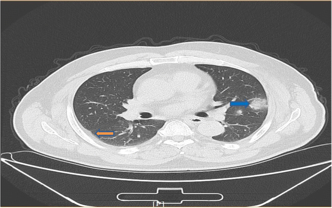
Objective: The outbreak of Severe Acute Respiratory Syndrome Coronavirus 2 (SARS-COV-2) has caused more than 26 million cases of Corona virus disease (COVID-19) in the world so far. To control the spread of the disease, screening large numbers of suspected cases for appropriate quarantine and treatment are a priority. Pathogenic laboratory testing is typically the gold standard, but it bears the burden of significant false negativity, adding to the urgent need of alternative diagnostic methods to combat the disease. Based on COVID-19 radiographic changes in CT images, this study hypothesized that artificial intelligence methods might be able to extract specific graphical features of COVID-19 and provide a clinical diagnosis ahead of the pathogenic test, thus saving critical time for disease control.
Methods: We collected 1065 CT images of pathogen-confirmed COVID-19 cases along with those previously diagnosed with typical viral pneumonia. We modified the inception transfer-learning model to establish the algorithm, followed by internal and external validation.
Results: The internal validation achieved a total accuracy of 89.5% with a specificity of 0.88 and sensitivity of 0.87. The external testing dataset showed a total accuracy of 79.3% with a specificity of 0.83 and sensitivity of 0.67. In addition, in 54 COVID-19 images, the first two nucleic acid test results were negative, and 46 were predicted as COVID-19 positive by the algorithm, with an accuracy of 85.2%.
Conclusion: These results demonstrate the proof-of-principle for using artificial intelligence to extract radiological features for timely and accurate COVID-19 diagnosis.
Key points: • The study evaluated the diagnostic performance of a deep learning algorithm using CT images to screen for COVID-19 during the influenza season. • As a screening method, our model achieved a relatively high sensitivity on internal and external CT image datasets. • The model was used to distinguish between COVID-19 and other typical viral pneumonia, both of which have quite similar radiologic characteristics.
Keywords: Artificial intelligence; COVID-19; Deep learning; Diagnosis; Tomography, X-ray computed.
© 2021. The Author(s).
Conflict of interest statement
The authors of this manuscript declare no relationships with any companies whose products or services may be related to the subject matter of the article.
Figures


References
MeSH terms
LinkOut - more resources
Full Text Sources
Other Literature Sources
Medical
Research Materials
Miscellaneous
