Review
. 2021 Jun;40(3):260-295.
doi: 10.1007/s10930-021-09967-8. Epub 2021 Feb 24.

A Biochemical Perspective of the Nonstructural Proteins (NSPs) and the Spike Protein of SARS CoV-2

Affiliations
Review

A Biochemical Perspective of the Nonstructural Proteins (NSPs) and the Spike Protein of SARS CoV-2

Francis K Yoshimoto. Protein J. 2021 Jun.

Abstract

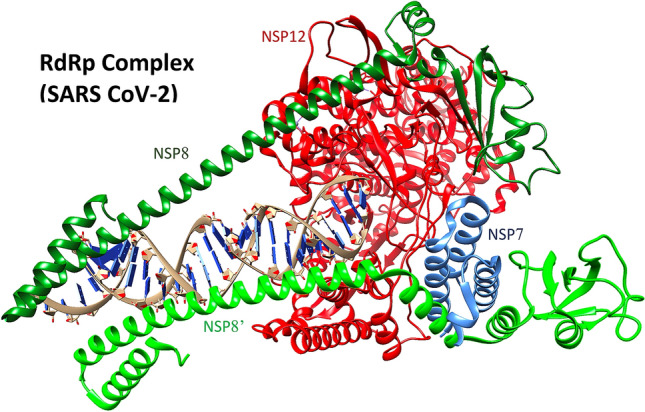
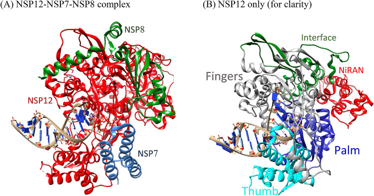
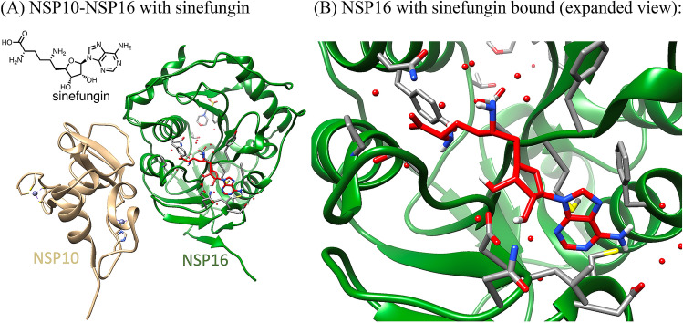
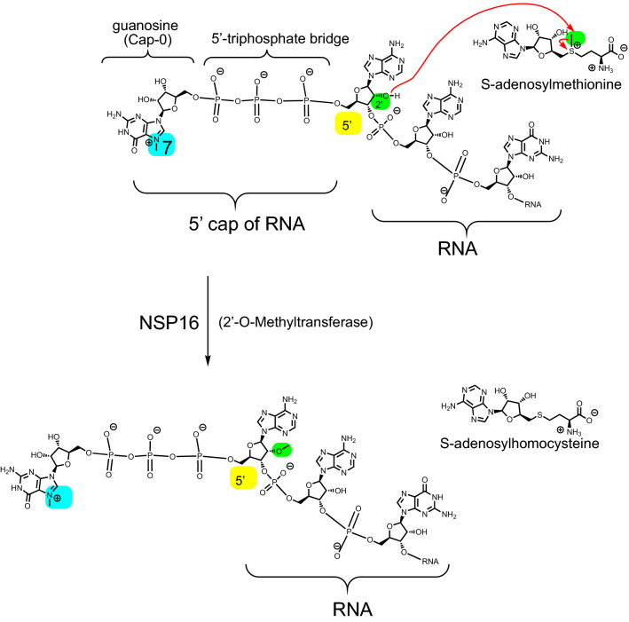
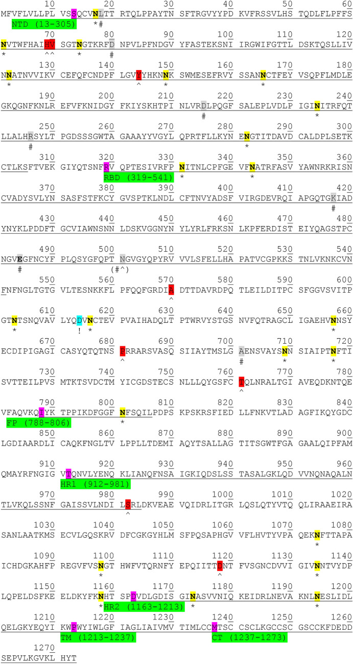
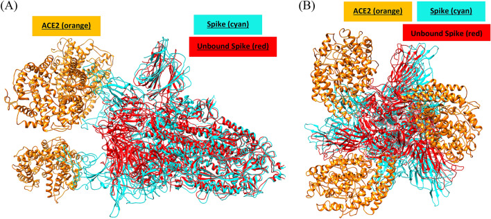
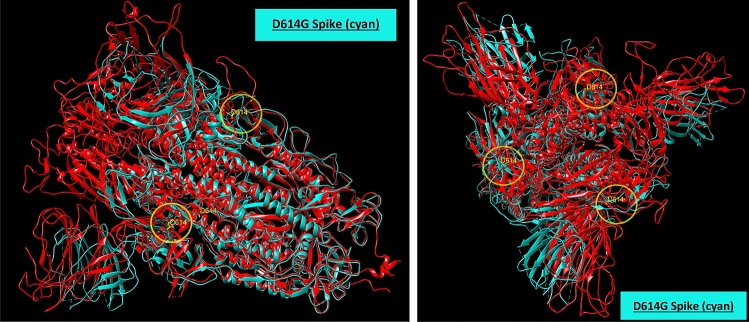
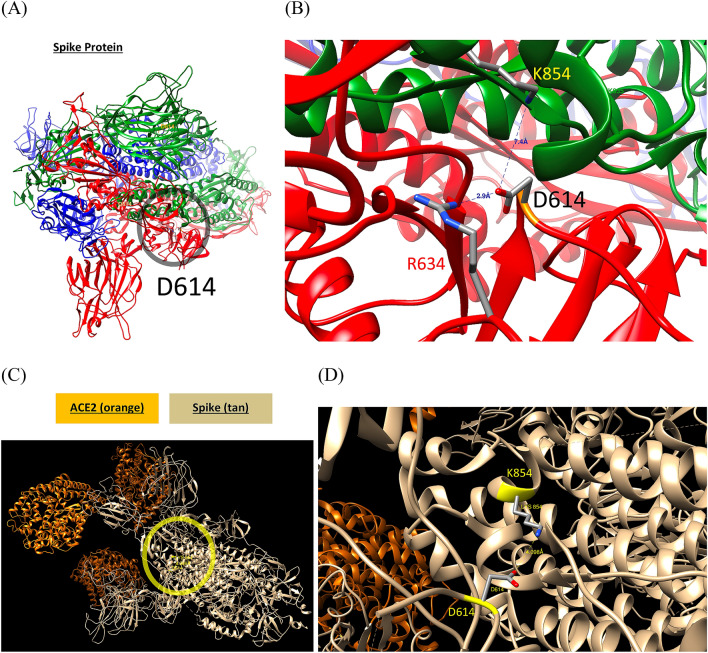
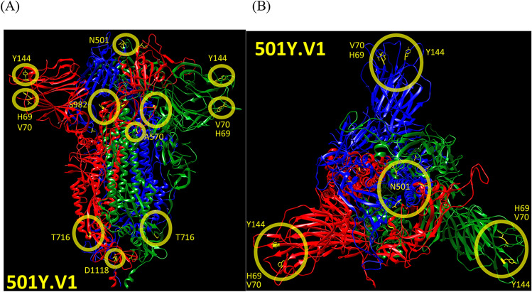
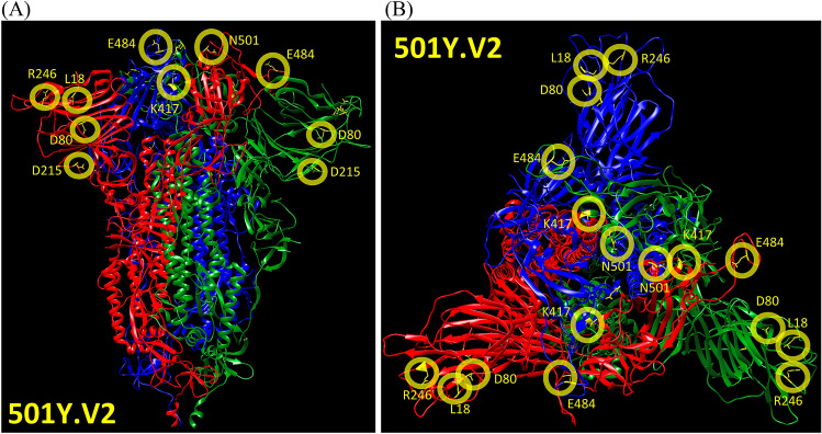
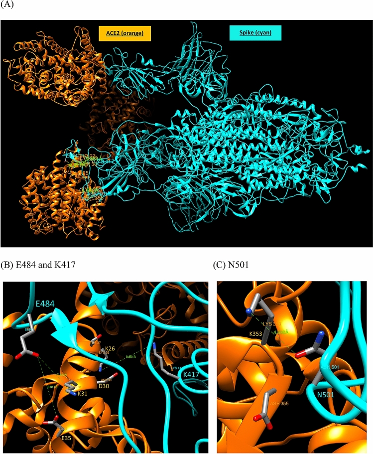
The global pandemic that shut down the world in 2020 was caused by the virus, SARS CoV-2. The chemistry of the various nonstructural proteins (NSP3, NSP5, NSP12, NSP13, NSP14, NSP15, NSP16) of SARS CoV-2 is discussed. Secondly, a recent major focus of this pandemic is the variant strains of SARS CoV-2 that are increasingly occurring and more transmissible. One strain, called "D614G", possesses a glycine (G) instead of an aspartate (D) at position 614 of the spike protein. Additionally, other emerging strains called "501Y.V1" and "501Y.V2" have several differences in the receptor binding domain of the spike protein (N501Y) as well as other locations. These structural changes may enhance the interaction between the spike protein and the ACE2 receptor of the host, increasing infectivity. The global pandemic caused by SARS CoV-2 is a rapidly evolving situation, emphasizing the importance of continuing the efforts to interrogate and understand this virus.

Keywords: Enzymes; Proteases; SARS CoV-2; Viral proteins.


Figures

Fig. 1
Fig. 1
The crystal structure of the papain like protase domain of NSP3 (PDB ID: 7CMD). Zoomed in region of the catalytic triad of NSP3 (aspartate-286, histidine-272, and cysteine-111)
Scheme 1
Scheme 1
The chemical reaction catalyzed by NSP3 (papain like protease or PLpro) and NSP5 (3C-like protease or 3CLpro). Also see Ref. [105] for more information. NSP3 has a catalytic triad (cysteine–histidine–aspartate) while NSP5 has a catalytic dyad (cysteine–histidine) [106]. For NSP3 the key residues are: D286, H272, and C111, and for NSP5, the active site residues are: H41 and C145
Fig. 2
Fig. 2
Crystal structure of the ADP ribose phosphatase domain of NSP3 (PDB ID: 6W02) [12]. The red spheres are water molecules (Color figure online)
Fig. 3
Fig. 3
a Crystal structure of NSP5 (PDB ID: 6Y2E). b Zoomed in view of the catalytic dyad (cysteine-145 and histidine-41) on the right (PDB ID: 6Y2E) [14]. c Crystal structure of NSP5 bound to inhibitor, GC-376 (PDB ID: 6WTT) [18]. d Zoomed in view of the catalytic dyad with inhibitor bound to the cysteine residue (C145). e Superimposed structures of (a) and (b). (apo protein is in red). f Zoomed in view of the superimposed structures. The distances between the histidine and the sulfur of the cysteine in the apo protein and inhibitor bound forms are 3.6 and 4.0 angstroms, respectively
Fig. 4
Fig. 4
Structure of the RNA-dependent RNA polymerase (RdRp) complex: NSP12 RNA polymerase (red) from SARS CoV-2 (PDB ID: 6YYT) [19]. The green proteins are the two NSP8 proteins (NSP8 and NSP8′) that are believed to interact and stabilize the RNA. NSP7 is shown in blue (Color figure online)
Fig. 5
Fig. 5
There are positively charged amino acid residues on NSP8 and NSP8′ (K37, K36, K40, K46, R51, R57, K58, K61) that stabilize the negatively charged phosphate groups in the RNA template (PDB ID: 6YYT) [19]
Scheme 2
Scheme 2
RNA polymerase reaction mechanism incorporating a new RNA (RNTP) into the primer strand
Fig. 6
Fig. 6
a Structure of NSP12 (red), also called: RNA-dependent RNA polymerase (RdRp) in complex with NSP7 (green) and NSP8 (blue) (PDB ID: 7BV2). b NSP12 alone with the RNA template (NSP7 and NSP8 are hidden for clarity). The different domains of NSP12 [98]—Nidovirus RdRp-associated nucleotidyltransferase (NiRAN): 51–249 (red), Interface: 250–365 (green), Fingers: 366–581 and 621–679 (grey), Palm: 582–620 and 680–815 (blue), and Thumb: 816–932 (cyan) (Color figure online)
Fig. 7
Fig. 7
The active site of NSP12 with remdesivir incorporated (PDB ID: 7BV2). The red sphere by C222 is a water molecule (Color figure online)
Fig. 8
Fig. 8
The metabolism of remdesivir into its triphosphate metabolite, the substrate of NSP12. Also shown is the structure of adenosine for comparison. The structure of favipiravir, another antiviral prodrug is shown
Fig. 9
Fig. 9
How remdesivir incorporation into the RNA primer inhibits RNA-dependent RNA polymerase activity (NSP12–NSP7–NSP8 complex) through chain termination. After incorporation of remdesivir into the primer strand, the RNA polymerase complex incorporates three more nucleotides before stalling. A hypothetical sequence for the template is shown above to illustrate that three NTPs are incorporated after remdesivir incorporation into the primer while the fourth NTP is not incorporated [20]
Fig. 10
Fig. 10
The structure of the 5′-cap of RNA, processed by the viral proteins of SARS CoV-2. The 5′-cap of viral RNA prevents recognition by the host innate immune system and promotes translation by the ribosome
Scheme 3
Scheme 3
The reaction catalyzed by NSP13 involving the RNA-5′-phosphatase activity to initiate the 5′-capping of mRNA. The amino acid residues K288 and D374 are proposed to play roles in promoting the terminal phosphate to leave and deprotonating the hydrolyzing water molecule, respectively. The support for this hypothesis is shown with the structure analysis in Fig. 11 (PDB ID: 6XEZ and 6YJT)
Fig. 11
Fig. 11
a Structural superposition between NSP13 of SARS CoV-2 and SARS CoV-1 (PDB ID: 6XEZ and 6YJT). Under the Matchmaker option in Chimera software, the reference chain was set to chain F (green) of 6XEZ (NSP13 complex of SARS CoV-2 PDB ID), and the chain to match was set to chain A (red) of 6YJT (PDB ID for NSP13 of SARS CoV-1, apo protein). b Focused view of the 5′-triphosphatase active site. c A different angle of SARS CoV-2 NSP13 (green, alone, PDB ID: 6XEZ) for clarity. d Expanded view of the active site of SARS CoV-2 NSP13 (green)—an AlF3 molecule is shown, which mimics the terminal monophosphate. The green spheres in b and d are Mg2+ ions (they are identical) (Color figure online)
Fig. 12
Fig. 12
The structure of NSP13 (grey) in complex with NSP7 (blue), NSP8 (green), and NSP12 (red) bound to an RNA template (PDB ID: 6XEZ). (There are two NSP13 units (NSP13 and NSP13′), two NSP8 units (NSP8 and NSP8′), one NSP12 unit, and one NSP7 unit) (Color figure online)
Fig. 13
Fig. 13
Structure of NSP13 (grey, helicase) in complex with NSP12 (red), NSP7 (blue), NSP8 (green), and RNA (PDB ID: 7XCM) [37]. (There are two NSP13 units (NSP13 and NSP13′), two NSP8 units (NSP8 and NSP8′), one NSP12 unit, and one NSP7 unit). NSP13′ has part of the RNA template bound, which shows the helicase activity of this protein (Color figure online)
Scheme 4
Scheme 4
The reaction catalyzed by NSP14 involving the N7-methylation of the guanosine residue of the 5′-cap of viral RNA. The methylating substrate is S-adenosylmethionine (SAM), which converts to S-adenosylhomocysteine (SAH)
Fig. 14
Fig. 14
Structure of NSP14 for SARS CoV-1 in complex with NSP10 (PDB ID: 5C8T). NSP14 in tan (right) and NSP10 is in red (left). An S-adenosylmethionine (SAM) ligand (green) is shown in the complex (circled). The green sphere is a magnesium (II) ion coordinated to the residues, D90 and E191 of NSP14 (Color figure online)
Scheme 5
Scheme 5
Endoribonuclease activity of NSP15
Fig. 15
Fig. 15
a Structure of apo NSP15 (green, PDB ID: 6VWW) [43]. b Structural alignment of NSP15 apo form (green, PDB ID: 6VWWL: 6VWW) and form bound to uridine diphosphate (red, PDB ID: 7K1O) [99]. c Structure of NSP15 from SARS CoV-2 bound to uridine diphosphate (red, PDB ID: 7K1O). d The expanded view of the active site of NSP15 with uridine diphosphate bound (PDB ID: 7K1O). The green spheres in a and b are water molecules (Color figure online)
Fig. 16
Fig. 16
a Structure of NSP10–NSP16 complex with sinefungin bound (PDB ID: 6YZ1). NSP16 is in green. NSP10 is tan. The structure of sinefungin is shown in the top left. b shows expanded view of the active site of NSP16 (green) with sinefungin (red) bound. The red spheres are water molecules (Color figure online)
Scheme 6
Scheme 6
The reaction catalyzed by NSP16. NSP16 transfers the methyl from S-adenosylmethionine to the 2′-O position in the 5′-cap of viral RNA
Fig. 17
Fig. 17
a Primary sequence alignment of SARS CoV-2 (GenBank: BCA87361.1) and SARS CoV-1 (GenBank: AAP13441.1). b The structural comparison of the spike proteins from SARS CoV-2 (red, PDB ID: 7JJJ) and SARS CoV-1 (light blue, PDB ID: 5XLR) [55]. AAP13441.1 (now obsolete but previously used: NP_828851.1 [1], where position S577A). c Rotated view (Color figure online)
Fig. 18
Fig. 18
a Primary sequence alignment of SARS CoV-2 (GenBank: BCA87361.1) and HCoV-229E (GenBank: QOP39313.1). b The structural comparison of the spike proteins from SARS CoV-2 (red, PDB ID: 7JJJ) and HCoV-229E (PDB ID: 6U7H) [52]. c Rotated view (Color figure online)
Fig. 19
Fig. 19
a Primary sequence alignment of SARS CoV-2 (GenBank: BCA87361.1) and MERS-CoV (GenBank: ASU91305.1). b The structural comparison of the spike proteins from SARS CoV-2 (red, PDB ID: 7JJJ) and MERS CoV (PDB ID: 5X5U, open RBD conformation) [56]. c Rotated view of (a) (Color figure online)
Fig. 20
Fig. 20
a Primary sequence alignment of SARS CoV-2 (GenBank: BCA87361.1) and HCoV OC43 (GenBank: AAA03055.1). b The structural comparison of the spike proteins from SARS CoV-2 (red, PDB ID: 7JJJ) and HCoV OC43 (PDB ID: 6OHW) [52]. c Rotated view of (a) (Color figure online)
Fig. 21
Fig. 21
a Primary sequence alignment of SARS CoV-2 (GenBank: BCA87361.1) and HCoV HKU1 (GenBank: ADN03339.1). b The structural comparison of the spike proteins from SARS CoV-2 (red, PDB ID: 7JJJ) and HCoV HKU1 (PDB ID: 5I08) [57]. c Rotated view (Color figure online)
Fig. 22
Fig. 22
a Primary sequence alignment of SARS CoV-2 (GenBank: BCA87361.1) and HCoV NL63 (GenBank: AGT51394.1). b The structural comparison of the spike proteins from SARS CoV-2 (red, PDB ID: 7JJJ) and HCoV NL63 (PDB ID: 5SZS) [58]. c Rotated view of (a) (Color figure online)
Fig. 23
Fig. 23
Structure of the spike protein trimer from SARS CoV-2 (PDB ID: 7JJJ). Each protomer is a different color (i.e. red, green, or blue) (Color figure online)
Fig. 24
Fig. 24
Structure of the spike protein (PDB ID: 7JJJ, chain a: red, chain b: blue, chain c: green) and highlighted are the different receptor binding domains (RBDs, position 319–541) for each protomer (green, blue, and red). a Side view (top) and rotated view (bottom) of the RBD of the spike protein. b The S1 fragment (position 14–685) of the spike protein is highlighted for each protomer (red, blue, and green) where the protease, furin, cleaves (side view, top) (rotated view, bottom), and c S2 fragment (686–1273) of the spike protein (side view, top) (rotated view, bottom) (Color figure online)
Fig. 25
Fig. 25
The sequence of the spike protein of SARS CoV-2. The 22 asparagine (N) residues that undergo glycosylation are highlighted. S1 subunit: 14–685 [100], S2 subunit: 686–1273, S2′ cleavage [61] site: 816. NTD N-terminal domain, RBD receptor binding domain, FP fusion peptide, HR1 heptapeptide repeat (or heptad repeat) sequence 1, HR2 heptad repeat 2, TM transmembrane domain, CT cytoplasm domain [101]. “*” indicates glycosylation sites. “!” Marks the location of the D614G variant (cyan) [47, 74]. “#” Marks the locations of the 501Y.V2 variant in grey ([i] NTD region: L18F, D80A, D215G, R246I, [ii] RBD region: K417N, E484K, N501Y, and [iii] A701V) [80]. “^” Marks the locations of the 501Y.V1 variant in red (H69, V70, Y144, (N501Y), A570D, P681H, T761I, S982A, D1118H–N501Y is already marked in grey with “#” for the 501Y.V2 variant) (Color figure online)
Fig. 26
Fig. 26
Structure of the spike protein (protomer) with asparagine residues that undergo glycosylation are highlighted in yellow (PDB ID: 7JJJ) (Color figure online)
Fig. 27
Fig. 27
Superimposed structures of (i) the spike protein with ACE2 bound (spike protein is cyan and ACE2 protein is orange, PDB ID: 7A98) and (ii) the unbound spike protein (red, PDB ID: 7JJJ). The open state of the spike protein can be seen when the ACE2 protein (orange) binds at the RBD of the spike protein (cyan). a Side view and b rotated view of (a) (Color figure online)
Fig. 28
Fig. 28
Illustration of SARS CoV-2 entry into the host cell via ACE2-B0AT1 (B0AT1 is also called SLC6A19, solute carrier family 6 member 19, sodium dependent neutral amino acid transporter) [102] (PDB ID: 6M18) [102]. (i) One receptor binding domain (RBD) (or two RBDs – PDB ID: 7A93) [70] of the spike protein orients in the open conformation (PDB ID: 6ZGG) [103] from the closed conformation (PDB ID: 6VXX) [60], (ii) ACE2 protein binds to the RBD of the spike protein (PDB ID: 7KNE) [104], (iii) a second RBD “opens” up (PDB ID: 7A96) [70], (iv) a second ACE2 protein binds to the second RBD of the spike protein (PDB ID: 7KMZ) [104], (v) a third ACE2 protein binds to the final RBD (PDB ID: 7KNI) [104], (vi) furin and TMPRSS2 cleave at the S1-S2 site and S2′ site of the spike protein releasing the ACE2-S1 complex (PDB ID: 7A92) [70] and in turn, leaving behind the S2 domain (PDB ID: 6XRA) [61], which is primed for entry into the host cell. Shown in the S2 domain trimer (PDB ID: 6XRA) is the spike protein sequence from T912-N1173 and Q1180-L1197. Interestingly, the spike protein with two RBD units in the open conformation has also been observed through cryo-EM (PDB ID: 7A93) [70] (Color figure online)
Fig. 29
Fig. 29
a Spike protein (cyan) bound to C105 neutralizing antibody Fab fragment (orange) (PDB ID: 6XCM) superimposed with spike protein (red, PDB ID: 7JJJ). b Rotated view of (a). c Spike protein (light blue) bound to S2A4 neutralizing antibody Fab fragment (green) (PDB ID: 7JVC) superimposed with spike protein (red, PDB ID: 7JJJ) (Color figure online)
Fig. 30
Fig. 30
Structural overlay of the spike protein D614 (red, PDB ID: 7JJJ) and G614 (cyan, PDB ID: 6XS6). Circled in yellow is the location of D614. The cryo-EM structure of the G614 variant has no RBD but is in a slightly more “open” conformation. a is the side view of the spike protein and b is the rotated view (Color figure online)
Fig. 31
Fig. 31
a A salt bridge between D614 and R634 within the same protomer (red) of the spike protein is shown. Another amino acid K854 from a different protomer (green) interacts with D614 (7.4 angstroms away) (PDB ID: 7JJJ). b is the zoomed in region of the salt bridge interactions (D614-R634 and D614-K854). The red color is one protomer and the green color is the second protomer (K854 is on a separate protomer from the D614 residue on the red protomer—they are 7.4 angstroms apart). Each protomer in the trimer is a different color: red, green, blue. c In the ACE2 bound-spike protein, D614, the K854 is shown closer to D614 (4.1 angstroms) suggesting a stabilizing role for this salt bridge (PDB ID: 7A98). d Zoomed in image of the salt bridge between D614 and K854 in the spike-ACE2 complex (4.1 angstroms apart) (Color figure online)
Fig. 32
Fig. 32
a The spike protein (PDB ID: 7JJJ) with the protomers colored differently (red, green, and blue). The amino acid residues that are changed in the 501Y.V1 variant are highlighted in yellow. b Rotated view. Highlighted in yellow are: H69 and V70 (deletion), Y144 (deletion), N501(Y), A570(D), T716(I), S982(A), and D1118(H)—position P681(H) is not included in the structure (Color figure online)
Fig. 33
Fig. 33
a Structure of the spike protein with the amino acid residue changes of the 501Y.V2 variant highlighted: L18F, D80A, D215G, R246I, K417N, E484K, N501Y, and A701V. b Rotated view (Color figure online)
Fig. 34
Fig. 34
a Spike protein-ACE2 protein complex (PDB ID: 7A98). Focus on the residues of the recently reported variant: K417N, E484K, and N501Y are shown in b and c. b E484 (spike) with K31 (ACE2) (the distance is also measured to E35 of the ACE2). c N501 (spike) with K353 (ACE2) is shown

Similar articles

Cited by

References

    1. Yoshimoto FK. The proteins of severe acute respiratory syndrome coronavirus-2 (SARS CoV-2 or n-COV19), the cause of COVID-19. Protein J. 2020;39:198–216. - PMC - PubMed
    1. Korber B, et al. Tracking changes in SARS-CoV-2 spike: evidence that D614G increases infectivity of the COVID-19 virus. Cell. 2020;182:812–827. - PMC - PubMed
    1. Walsh EE, et al. Safety and immunogenicity of two RNA-based Covid-19 vaccine candidates. NEJM. 2020;383:2439. - PMC - PubMed
    1. Mulligan MJ, et al. Phase I/II study of COVID-19 RNA vaccine BNT162b1 in adults. Nature. 2020;585:589–593. - PubMed
    1. Jackson LA, et al. An mRNA vaccine against SARS-CoV-2 - perliminary report. NEJM. 2020;383:1920–1931. - PMC - PubMed

MeSH terms

Substances