. 2021 May;31(3):e12936.
doi: 10.1111/bpa.12936. Epub 2021 Feb 24.

Distinct and dementia-related synaptopathy in the hippocampus after military blast exposures

Affiliations

Distinct and dementia-related synaptopathy in the hippocampus after military blast exposures

Michael F Almeida et al. Brain Pathol. 2021 May.

Abstract

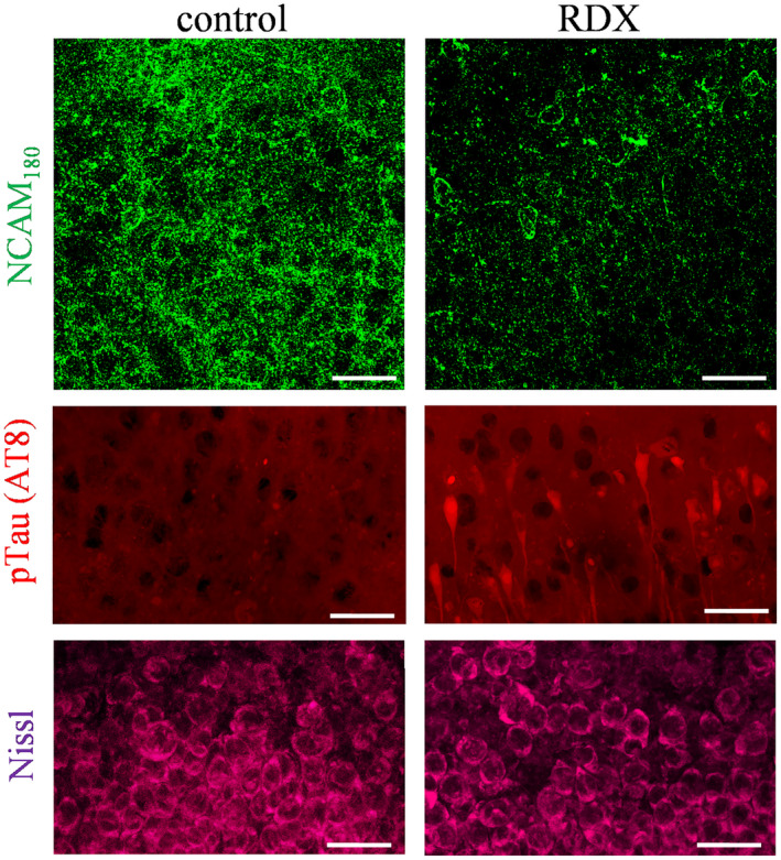
Explosive shockwaves, and other types of blast exposures, are linked to injuries commonly associated with military service and to an increased risk for the onset of dementia. Neurological complications following a blast injury, including depression, anxiety, and memory problems, often persist even when brain damage is undetectable. Here, hippocampal explants were exposed to the explosive 1,3,5-trinitro-1,3,5-triazinane (RDX) to identify indicators of blast-induced changes within important neuronal circuitries. Highly controlled detonations of small, 1.7-gram RDX spherical charges reduced synaptic markers known to be downregulated in cognitive disorders, but without causing overt neuronal loss or astroglial responses. In the absence of neuromorphological alterations, levels of synaptophysin, GluA1, and synapsin IIb were significantly diminished within 24 hr, and these synaptic components exhibited progressive reductions following blast exposure as compared to their stable maintenance in control explants. In contrast, labeling of the synapsin IIa isoform remained unaltered, while neuropilar staining of other markers decreased, including synapsin IIb and neural cell adhesion molecule (NCAM) isoforms, along with evidence of NCAM proteolytic breakdown. NCAM180 displayed a distinct decline after the RDX blasts, whereas NCAM140 and NCAM120 exhibited smaller or no deterioration, respectively. Interestingly, the extent of synaptic marker reduction correlated with AT8-positive tau levels, with tau pathology stochastically found in CA1 neurons and their dendrites. The decline in synaptic components was also reflected in the size of evoked postsynaptic currents recorded from CA1 pyramidals, which exhibited a severe and selective reduction. The identified indicators of blast-mediated synaptopathy point to the need for early biomarkers of explosives altering synaptic integrity with links to dementia risk, to advance strategies for both cognitive health and therapeutic monitoring.

Keywords: Alzheimer-type synaptopathogenesis; NBDP; NCAM breakdown products; mild traumatic brain injury; neurotrauma; synaptic decline.


Conflict of interest statement

The authors declare that they have no competing interests. The funding agencies had no role in study design, data collection and analysis, or decision to publish.

Figures

FIGURE 1
FIGURE 1
Military blast‐generated shockwaves lead to synaptic alteration in hippocampal explants. (A) Transverse slices of rat hippocampi were maintained on 3‐cm inserts for 18‐22 days in a six‐well culture plate. (B) The culture plate was sealed in serum‐free medium and clamped in a water‐filled chamber for military blast exposures. Control slice cultures exposed to the serum‐free conditions of the apparatus were assessed by Nissl (C) and synaptophysin staining (D), showing the stable maintenance of hippocampal subfields and associated dense neuropil (view‐field width: 1.5 mm). (E) Small spherical assemblies of RDX explosive were detonated outside the tank. (F) The shockwave propagated into the tank and was recorded as a time‐pressure profile by three sensors located above the culture plate. (G) Explants subjected to RDX detonations were assessed by immunoblotting for synaptophysin levels (SNP) and compared to control explants treated with identical conditions without blasts (con) or with mechanical stress produced by consecutive flexing protocols applied to the Biopore insert membrane. (H) Control and blast‐exposed explant cultures were fixed and subjected to synaptophysin, synapsin IIa, and Nissl staining (scale bars: 50 µm). sp, stratum pyramidale; sr; stratum radiatum
FIGURE 2
FIGURE 2
Blast waves from RDX spherical charges induce distinct synaptic vulnerability. (A) Hippocampal explants were subjected to three consecutive RDX detonations, then maintained in culture for 24‐45 hr, and subsequently assessed for GluA1 (n = 8), synaptophysin (SNP; n = 8), synaptotagmin‐5 (Syt‐5; n = 4), synapsin IIa (syn IIa; n = 6) and IIb (syn IIb; n = 9), and actin (n = 7) as a load control, with comparisons to explants treated with identical conditions without blasts (0 hr). (B) For the immunostained proteins, integrated optical densities were measured to determine blast‐induced changes as compared to respective measures from control explants (means ± SEM). *p < 0.05; ***p < 0.001. (C and D) Across post‐blast times, mean immunoreactivities were normalized to respective data from three control sets of pooled explants that received identical conditions without blasts and harvested 45 hr later. The following results were from one‐way ANOVA tests: SNP, p = 0.0001 (n = 11); GluA1, p < 0.0001 (n = 11); syn IIb, p < 0.0001 (n = 7). (E) Slice cultures treated with or without RDX blasts were fixed and subjected to synaptophysin and GluA1 immunolabeling, followed by DAPI counterstaining (scale bars: 50 µm). sp, stratum pyramidale; sr; stratum radiatum
FIGURE 3
FIGURE 3
NCAM isoforms exhibit distinct blast‐induced effects. (A) Three NCAM isoforms were assessed in cultured hippocampal slices that were exposed to consecutive RDX detonations and compared to control explants treated identically without blasts (con). The immunoblots were also analyzed with longer chemiluminescent exposure times, revealing NCAM breakdown products (NBDPs, red boxes). (B) The NCAM isoforms were selectively labeled by anti‐NCAM antibody as there was no detectable labeling of 120‐, 140‐, or 180‐kDa proteins in the blot strip incubated with non‐selective IgG during the immunoblotting protocol. (C) NCAM immunoreactivity measures (n = 7) were compared to their respective levels in control explants (mean percent change ± SEM), and the blast‐induce effects were determined to be significantly different among the isoforms (Kruskal‐Wallis test: p < 0.001)
FIGURE 4
FIGURE 4
Blast‐induced reduction of neuropilar NCAM180 labeling was associated with pathological tau staining. Blast‐exposed and control explants were fixed and immunolabeled for the vulnerable NCAM180 isoform (top panels; scale bar: 25 µm) and for phosphorylated tau (pTau) using the AT8 monoclonal antibody (middle panels; scale bar: 50 µm). Counterstaining by Nissl protocol found no obvious indications of neuronal alterations (bottom panels; scale bar: 25 µm). sp, stratum pyramidale; sr; stratum radiatum
FIGURE 5
FIGURE 5
Blast‐induced functional changes in CA1 neurons. (A) Hippocampal slice cultures were subjected to detonations of 1‐3 RDX spherical charges then assessed 24‐48 hr later using whole‐cell patch clamp to record intrinsic properties. (B) Control explants that received identical conditions without blasts (con; n = 20) and the RDX‐treated explants (n = 11) were assessed for resting membrane potential in pyramidal neurons (RMP; means ± SEM). (C) In control (n = 19) and blast‐exposed explants (n = 12), the input resistance was also measured in the neurons (one‐way ANOVA: p = 0.0013; Bonferroni test: **p < 0.001). (D) Additional measures found that action potential firing threshold was unchanged across the explant treatment groups (left graph), while action potential half‐width exhibited a 45% increase after multiple blasts compared to controls (right graph; ANOVA p = 0.0157; Bonferroni test: *p = 0.014). (E) Representative traces are shown for control explants and those exposed to three consecutive blasts. (F) The blast exposures reduced the amplitude of evoked postsynaptic currents (PSC) as well in response to varied stimulation intensities to the Schaffer collateral inputs. (G) Note the 10‐fold difference in amplitude scales between the two sets of representative traces. (H) A subset of the control explants (n = 6) and the RDX‐exposed explants (n = 12) were subsequently stained for synaptophysin, GFAP, and DAPI (scale bars: 50 µm). (I) Resulting from image analysis of confocal micrographs, synaptophysin immunostaining in CA1 revealed blast‐induced progressive decline in the neuropil (ANOVA p < 0.01; Tukey test: **p = 0.0057). (J) In the same assessed area of the stratum radiatum, GFAP immunostaining showed no indication of early astroglial activation by area fraction analysis. sp, stratum pyramidale; sr, stratum radiatum

Similar articles

Cited by

References

    1. Brix KA, Brody DL, Grimes JB, Yitzhak A. Working Group Members . Military blast exposure and chronic neurodegeneration: summary of working groups and expert panel findings and recommendations. J Neurotrauma 2017;34(S1):S18–25.
    1. Department of Defense (DoD) . Data from “Worldwide numbers for traumatic brain injury”. https://dvbic.dcoe.mil/dod‐worldwide‐numbers‐tbi. Accessed May 15, 2020.
    1. Williamson V, Mulhall E. Invisible wounds: psychological and neurological injuries confront a new generation of veterans. Iraq and Afghanistan Veterans of America, Issue Report; 2009.
    1. Goldstein LE, Fisher AM, Tagge CA, Zhang X‐L, Velisek L, Sullivan JA, et al. Chronic traumatic encephalopathy in blast‐exposed military veterans and a blast neurotrauma mouse model. Sci Transl Med. 2012;4:134ra60. - PMC - PubMed
    1. Sawyer TW, Ritzel DV, Wang Y, Josey T, Villanueva M, Nelson P, et al. Primary blast causes delayed effects without cell death in shell‐encased brain cell aggregates. J Neurotrauma. 2018;35:174–86. - PubMed

Publication types

Substances