

Histological and immunohistochemical changes in facial skin treated with combined ablative and non-ablative laser therapy
- PMID: 33629512
- PMCID: PMC8597027
- DOI: 10.1111/jocd.14023
Histological and immunohistochemical changes in facial skin treated with combined ablative and non-ablative laser therapy
Abstract
Background: Facial skin rejuvenation is a highly sought after procedure. Different materials, energy-based devices and techniques have been shown to offer good results in facial rejuvenation.
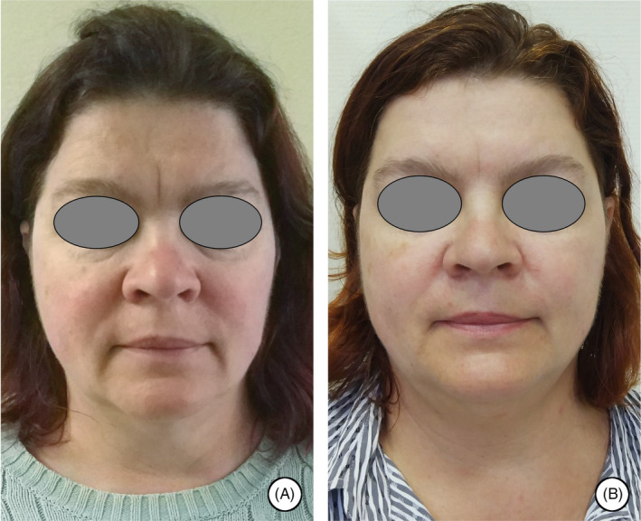
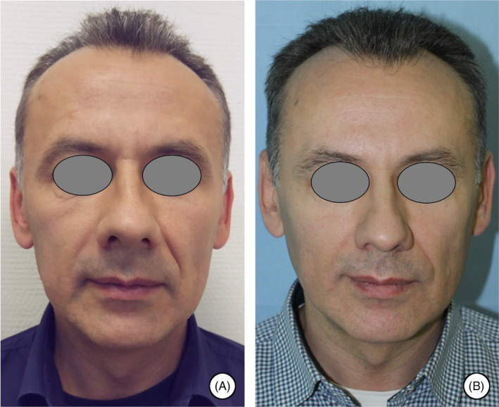
Aims: The aim of this study was to evaluate the macroscopic and histological and immunohistochemical changes in facial skin treated with a combined laser therapy.
Patients/methods: Fourteen patients aged from 38 to 59 years were included in the study. Two different wavelengths (2940 and 1064 nm) were used in this four-step procedure.
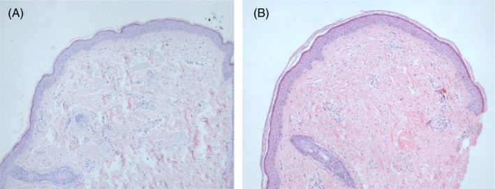
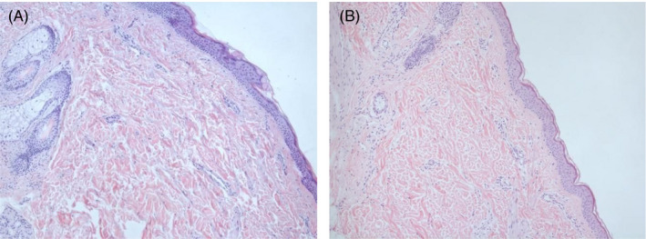
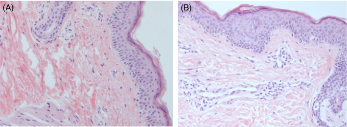
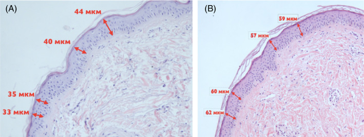
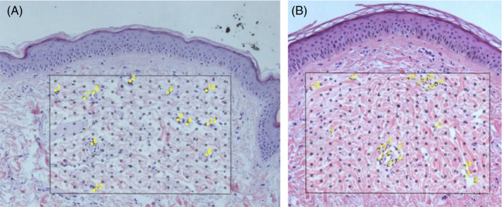
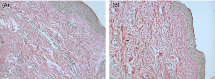
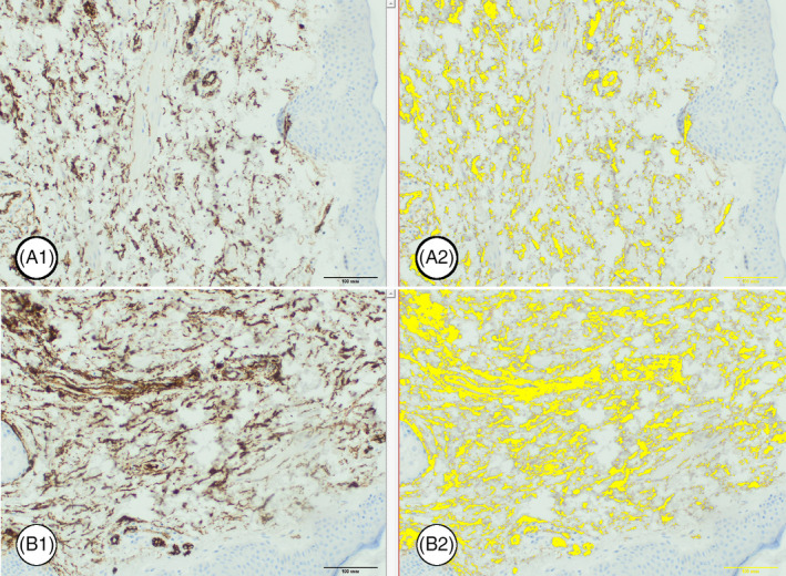
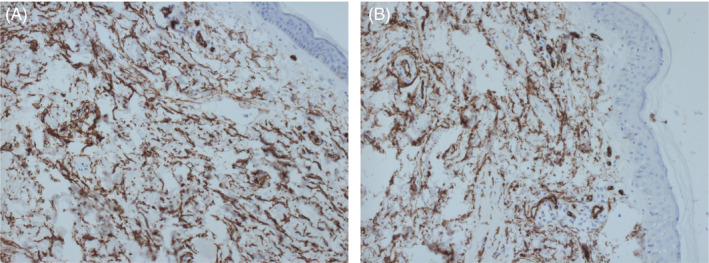
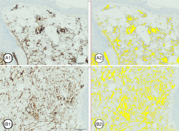
Results: Significant improvement according to classification for age-related changes in all areas of the face was achieved. There were also significant structural changes in the epidermis and dermis, confirmed with histology and immunohistochemistry.
Conclusion: The dual-wavelength protocol has been found to provide excellent results for facial rejuvenation.
Keywords: 4D; Er:YAG; Nd:YAG; facial rejuvenation; histology.
© 2021 The Authors. Journal of Cosmetic Dermatology published by Wiley Periodicals LLC.
Conflict of interest statement
All authors state that they have no conflicts of interest regarding this article.
Figures
Similar articles
-
Multifractional microablative laser combined with spacially modulated ablative (SMA) technology for facial skin rejuvenation.Lasers Surg Med. 2017 Jan;49(1):78-83. doi: 10.1002/lsm.22561. Epub 2016 Jul 18. Lasers Surg Med. 2017. PMID: 27426249
-
Combined fractional ablative and nonablative laser resurfacing treatment: a split-face comparative study.J Drugs Dermatol. 2013 Feb;12(2):175-8. J Drugs Dermatol. 2013. PMID: 23377390
-
Delivery of light to the skin through ablated conduits.Lasers Surg Med. 2017 Jan;49(1):69-77. doi: 10.1002/lsm.22533. Epub 2016 May 19. Lasers Surg Med. 2017. PMID: 27197620 Review.
-
Fractional versus ablative erbium:yttrium-aluminum-garnet laser resurfacing for facial rejuvenation: an objective evaluation.J Am Acad Dermatol. 2013 Jan;68(1):103-12. doi: 10.1016/j.jaad.2012.09.014. Epub 2012 Oct 27. J Am Acad Dermatol. 2013. PMID: 23110966
-
Lasers for facial rejuvenation.Am J Clin Dermatol. 2003;4(4):225-34. doi: 10.2165/00128071-200304040-00002. Am J Clin Dermatol. 2003. PMID: 12680801 Review.
Cited by
-
Non-ablative Er:YAG laser is an effective tool in the treatment arsenal of androgenetic alopecia.J Cosmet Dermatol. 2022 May;21(5):2056-2063. doi: 10.1111/jocd.14370. Epub 2021 Aug 26. J Cosmet Dermatol. 2022. PMID: 34435735 Free PMC article.
-
Comparative analysis of erbium: glass 1550 nm and combined erbium: YAG & Nd: YAG lasers for perioral rejuvenation: a prospective study.Lasers Med Sci. 2025 Jun 20;40(1):291. doi: 10.1007/s10103-025-04540-6. Lasers Med Sci. 2025. PMID: 40537663 Clinical Trial.
-
Hair regrowth and maintenance in alopecia universalis patient treated with nonablative Er:YAG laser.Clin Case Rep. 2021 Nov 12;9(11):e04948. doi: 10.1002/ccr3.4948. eCollection 2021 Nov. Clin Case Rep. 2021. PMID: 34804525 Free PMC article.
-
Concomitant Use of Botulinum Toxin and Super Long Nd:YAG Laser.J Cosmet Dermatol. 2025 Mar;24(3):e70042. doi: 10.1111/jocd.70042. J Cosmet Dermatol. 2025. PMID: 40052648 Free PMC article.
-
Keloid and Hypertrophic Scars Treatment.Aesthetic Plast Surg. 2024 Jul;48(13):2553-2560. doi: 10.1007/s00266-024-03869-7. Epub 2024 Mar 7. Aesthetic Plast Surg. 2024. PMID: 38453710
References
-
- Matts PJ, Fink B, Grammer K, Burquest M. Color homogeneity and visual perception of age, health, and attractiveness of female facial skin. J Am Acad Dermatol. 2007;57(6):977‐984. - PubMed
-
- Sadick NS, Cardona A. Laser treatment for facial acne scars: a review. J Cosmet Laser Ther. 2018;20(7–8):424‐435. - PubMed
-
- Fitzpatrick RE, Rostan EF, Marchell N. Collagen tightening induced by carbon dioxide laser versus erbium: YAG laser. Lasers Surg Med. 2000;27(5):395‐403. - PubMed
MeSH terms
LinkOut - more resources
Full Text Sources
Other Literature Sources
Medical
