

A freeze-and-thaw-induced fragment of the microtubule-associated protein tau in rat brain extracts: implications for the biochemical assessment of neurotoxicity
- PMID: 33629708
- PMCID: PMC7990086
- DOI: 10.1042/BSR20203980
A freeze-and-thaw-induced fragment of the microtubule-associated protein tau in rat brain extracts: implications for the biochemical assessment of neurotoxicity
Abstract
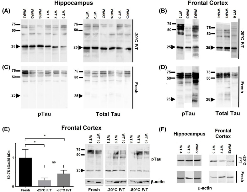
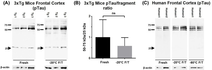
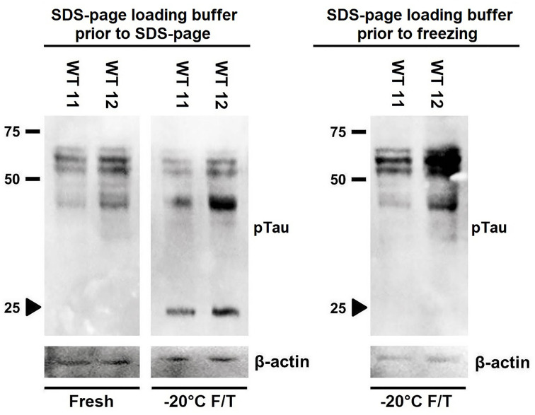
Tau is a microtubule-associated protein (MAP) responsible for controlling the stabilization of microtubules in neurons. Tau function is regulated by phosphorylation. However, in some neurological diseases Tau becomes aberrantly hyperphosphorylated, which contributes to the pathogenesis of neurological diseases, known as tauopathies. Western blotting (WB) has been widely employed to determine Tau levels in neurological disease models. However, Tau quantification by WB should be interpreted with care, as this approach has been recognized as prone to produce artifactual results if not properly performed. In the present study, our goal was to evaluate the influence of a freeze-and-thaw cycle, a common procedure preceding WB, to the integrity of Tau in brain homogenates from rats, 3xTg-AD mice and human samples. Homogenates were prepared in ice-cold RIPA buffer supplemented with protease/phosphatase inhibitors. Immediately after centrifugation, an aliquot of the extracts was analyzed via WB to quantify total and phosphorylated Tau levels. The remaining aliquots of the same extracts were stored for at least 2 weeks at either -20 or -80°C and then subjected to WB. Extracts from rodent brains submitted to freeze-and-thaw presented a ∼25 kDa fragment immunoreactive to anti-Tau antibodies. An in-gel digestion followed by mass spectrometry (MS) analysis in excised bands revealed this ∼25 kDa species corresponds to a Tau fragment. Freeze-and-thaw-induced Tau proteolysis was detected even when extracts were stored at -80°C. This phenomenon was not observed in human samples at any storage condition tested. Based on these findings, we strongly recommend the use of fresh extracts of brain samples in molecular analysis of Tau levels in rodents.
Keywords: Alzheimer's disease; disease models; neurodegeneration; tau protein; tauopathy; western blot.
© 2021 The Author(s).
Conflict of interest statement
The authors declare that there are no competing interests associated with the manuscript.
Figures
Similar articles
-
Western Blot of Tau Protein from Mouse Brains Extracts: How to Avoid Signal Artifacts.Methods Mol Biol. 2024;2754:309-321. doi: 10.1007/978-1-0716-3629-9_16. Methods Mol Biol. 2024. PMID: 38512673
-
Identification of high-performing antibodies for the reliable detection of Tau proteoforms by Western blotting and immunohistochemistry.Acta Neuropathol. 2024 May 18;147(1):87. doi: 10.1007/s00401-024-02729-7. Acta Neuropathol. 2024. PMID: 38761203 Free PMC article.
-
A NH2 tau fragment targets neuronal mitochondria at AD synapses: possible implications for neurodegeneration.J Alzheimers Dis. 2010;21(2):445-70. doi: 10.3233/JAD-2010-100120. J Alzheimers Dis. 2010. PMID: 20571215
-
Tauopathy: A common mechanism for neurodegeneration and brain aging.Mech Ageing Dev. 2019 Mar;178:72-79. doi: 10.1016/j.mad.2019.01.007. Epub 2019 Jan 19. Mech Ageing Dev. 2019. PMID: 30668956 Free PMC article. Review.
-
Tau as a therapeutic target for Alzheimer's disease.Curr Alzheimer Res. 2011 Sep;8(6):666-77. doi: 10.2174/156720511796717195. Curr Alzheimer Res. 2011. PMID: 21679154 Free PMC article. Review.
Cited by
-
Comprehensive review of drug resistance in mammalian cancer stem cells: implications for cancer therapy.Cancer Cell Int. 2024 Dec 18;24(1):406. doi: 10.1186/s12935-024-03558-0. Cancer Cell Int. 2024. PMID: 39695669 Free PMC article. Review.
References
-
- Jeganathan S., Hascher A., Chinnathambi S., Biernat J., Mandelkow E.-M. and Mandelkow E. (2008) Proline-directed pseudo-phosphorylation at AT8 and PHF1 epitopes induces a compaction of the paperclip folding of Tau and generates a pathological (MC-1) conformation. J. Biol. Chem. 283, 32066–32076 10.1074/jbc.M805300200 - DOI - PubMed
Publication types
MeSH terms
Substances
LinkOut - more resources
Full Text Sources
Other Literature Sources
Medical
Molecular Biology Databases