Review
. 2021 May;78(9):4305-4333.
doi: 10.1007/s00018-021-03787-w. Epub 2021 Feb 25.

Ubiquitin-related processes and innate immunity in C. elegans

Affiliations
Review

Ubiquitin-related processes and innate immunity in C. elegans

Juan A Garcia-Sanchez et al. Cell Mol Life Sci. 2021 May.

Abstract

Innate immunity is an evolutionary ancient defence strategy that serves to eliminate infectious agents while maintaining host health. It involves a complex network of sensors, signaling proteins and immune effectors that detect the danger, then relay and execute the immune programme. Post-translational modifications relying on conserved ubiquitin and ubiquitin-like proteins are an integral part of the system. Studies using invertebrate models of infection, such as the nematode Caenorhabditis elegans, have greatly contributed to our understanding of how ubiquitin-related processes act in immune sensing, regulate immune signaling pathways, and participate to host defence responses. This review highlights the interest of working with a genetically tractable model organism and illustrates how C. elegans has been used to identify ubiquitin-dependent immune mechanisms, discover novel ubiquitin-based resistance strategies that mediate pathogen clearance, and unravel the role of ubiquitin-related processes in tolerance, preserving host fitness during pathogen attack. Special emphasis is placed on processes that are conserved in mammals.

Keywords: Host–pathogen interaction; Proteostasis; SUMOylation; Ubiquitination; Unfolded protein response.


Conflict of interest statement

The authors declare that they have no competing interests. All authors contributed to the writing of the manuscript.

Figures

Fig. 1
Fig. 1
Regulation and functional consequences of the ubiquitin reaction. a Amino-acid sequence of human ubiquitin. Red: the N-terminal methionine and the seven lysine residues are acceptor sites that can be modified by the addition of another ubiquitin molecule. Blue: glycine 76 allows covalent linking to a substrate or another ubiquitin molecule. b Ubiquitination diversity and recognition. Ubiquitinated substrates can bind with a variety of ubiquitin binding proteins (UBPs) that possess ubiquitin-binding domains recognizing the different ubiquitination types. UBPs binding mixed or branched chains remain to be identified. A single ubiquitin molecule can attach to a substrate via one (mono-Ub) or several (multi-Ub) lysine residues. In addition, the first methionine and/or one of the seven lysine residues of ubiquitin can serve as ubiquitin acceptor sites, forming poly-Ub chains with different topologies. Homotypic poly-Ub chains, engaging the same linkage within the polymer, can display a compact (K48) or linear (K63 and M1) conformation. Heterotypic poly-Ub chains include chains with mixed linkages or branched chains, with two distal ubiquitin molecules attached to at least two acceptor sites of a single proximal ubiquitin moiety. c Ubiquitin reaction and outcome. E1 activates Ub in an ATP-dependent manner, then transfers Ub to an E2 Ub-conjugating enzyme. The E2 active site cysteine forms a thioester bond (represented as ~) with the C-terminal carboxyl group of Ub. Finally, E3 Ub-ligases mediate the transfer of Ub from the E2 to a lysine residue of the substrate. De-ubiquitinating enzymes (DUBs) can reverse the reaction, restoring the pool of free ubiquitin. K48 and K11 chains, which display a compact conformation, are known to direct proteasomal degradation, while K63 and M1 linear chains are involved in autophagy and intracellular signaling
Fig. 2
Fig. 2
Tripartite structure of E3-CRL. a Schematic representation of Cullin-RING E3 ligase (E3-CRL) multimeric complexes. A catalytic RING-containing enzyme binds to the E2 ~ Ub intermediate and the cullin subunit, which acts as a scaffold of the complex. Interaction with the substrate is mediated by a substrate recognition subunit (SRS) that interacts with the cullin subunit either directly or indirectly via an adaptor protein. E3-CRLs mediate the direct transfer of Ub from the E2 to the substrate (dashed arrow). b SRS and adaptor families. BTB (Broad-complex, Tramtrack and Bric-à-brac) containing SRSs bind directly to the cullin subunit. F-box, SOCS-BC (Suppressor of Cytokine Signaling protein 1, binding to EloB-C) and DCAF (DDB1-CUL4-Associated Factor)-containing SRSs interact with cullin via SKR (Skp-1 Related), EloB-C (Elongin B-C complex) and DDB1 (DNA damage-binding protein 1), respectively
Fig. 3
Fig. 3
Model of Ub-dependent mechanisms in surveillance immunity against microsporidia. Intracellular microsporidia cells interfere with proteasome function through an unknown mechanism. This triggers surveillance immunity and induction of “intracellular pathogen response” genes such as cul-6, skr-3, skr-4 and skr-5. CUL-6-dependent E3-CRL then promotes proteostasis on one hand, and ubiquitination and subsequent elimination of microsporidian cells on the other. Together, CUL-6-mediated proteostasis and pathogen ubiquitination contributes to host defence
Fig. 4
Fig. 4
Model of neuronal regulation of NPR-1 by the E3-HECT ligase HECW-1. a Respective position of OLL (yellow) and RMG (red) neurons in the anterior and posterior bulb of the pharynx (green). Adapted from WormAtlas (https://www.wormatlas.org/) b Schematic intercellular signaling pathway of P. aeruginosa sensing and subsequent induction of avoidance behaviour. During infection, HECW-1, which is expressed in OLL sensory neurons, must induce ubiquitination of a yet to identify substrate. This regulation ultimately inhibits activity of the NPR-1 receptor localized in RMG neurons, impairing avoidance behaviour
Fig. 5
Fig. 5
Model of PMK-1 pathway regulation by Ub-related genes during P. aeruginosa and D. coniospora infection. D. coniospora activates the PMK-1 pathway following binding of HPLA to the GPCR DCAR-1. This triggers expression of genes such as nlp-29 encoding antimicrobial peptides, via the transcription factor STA-2. Seven Ub-modifying enzymes and three proteasomal subunits potentially positively regulate the DCAR-1/PMK-1/STA-2 pathway in the epidermis (green box #1). The transcription co-factor AKIR-1, which binds to the POU transcription factor CEH-18 and regulates expression of nlp-29 in the epidermis upon fungal infection, also interacts with and might be regulated by the Ub-related enzymes UBR-5, MATH-33 and USP-24. In intestinal cells, through an uncharacterised mechanism, P. aeruginosa also activates the PMK-1 pathway, and triggers expression of genes such as clec-85 that encode antimicrobial proteins, through the transcription factor ATF-7. Three Ub-modifying enzymes are potential positive regulators of this pathway (green box #2), including SIAH-1 which may directly regulate TIR-1. RLE-1 is a negative regulator of the pathway, specifically targeting NSY-1 to the UPS (red box #3)
Fig. 6
Fig. 6
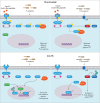
Model of Ub-dependent regulation of the IIS signaling pathway. Under conditions where IIS is activated, the receptor DAF-2 auto-phosphorylates and activates downstream kinases, leading to the phosphorylation of DAF-16 and its retention in the cytosol by 14-3-3 (upper panels). In condition of low IIS, unphosphorylated DAF-16 is targeted to the nucleus and enhances host defence against P. aeruginosa and Cry5B pore-forming toxin (PFT) (lower panels). The DUB MATH-33 is also targeted to the nucleus of intestinal cells, and interacts, deubiquitinates and stabilizes DAF-16, boosting host defence against P. aeruginosa (left panel). It is not known what E3 ligase ubiquitinates DAF-16 nor whether this occurs in the cytoplasm or in the nucleus. In parallel, the E3-HECT ligase WWP-1 acts downstream of PDK-1 and enhances host defence against PFT, probably through the Ub-dependent regulation of one or several substrates. PDK-1 may regulate WWP-1 through an inhibitory phosphorylation
Fig. 7
Fig. 7
Model of Ub and Ubl-dependent regulation of mitochondrial damage responses during P. aeruginosa infection. Depending on the culture conditions, P. aeruginosa virulence involves pyoverdine (left) or cyanide and phenazines (right), virulence factors that induce mitochondrial damage. Following pyoverdine exposure, damaged mitochondria are cleared by mitophagy in a PDR-1-dependent manner, which provides protection against P. aeruginosa infection. By analogy with mammalian autophagy, PDR-1 might induce ubiquitination of a mitochondrial outer protein, which might recruit UBP bound to the isolation membrane to promote mitochondrial engulfment into autophagosome. Fusion of autophagosomes with lysosomes induces degradation of damaged mitochondria in autophagolysosome structures in a process known as mitophagy. In solid culture, mitochondrial damage leads to increased expression of the deSUMOylase ULP-4, which removes the SUMO homologue SMO-1 from ATFS-1 and DVE-1. DeSUMOylated ATFS-1 displays increased stability and transcriptional activity, while deSUMOylation of DVE-1 increases its nuclear translocation. The increased expression of innate immune genes and/or UPRmt ultimately promotes host defence. P. aeruginosa production of phenazines represses UPRmt in a ZIP-3 dependent manner. ZIP-3 negatively regulates ATFS-1-mediated host defence. WWP-1, induced upon mitochondrial stress, appears to be the E3 ligase responsible for ZIP-3 degradation by UPS. Data suggest a model where P. aeruginosa prevents ZIP-3 UPS degradation to counteract ATFS-1 mediated host defence. Although shown here in the cytoplasm, the site of ZIP-3 UPS regulation is not known

Similar articles

Cited by

References

    1. Medzhitov R. Approaching the asymptote: 20 Years Later. Immunity. 2009;30:766–775. doi: 10.1016/j.immuni.2009.06.004. - DOI - PubMed
    1. Curtis VA. Infection-avoidance behaviour in humans and other animals. Trends Immunol. 2014;35:457–464. doi: 10.1016/j.it.2014.08.006. - DOI - PubMed
    1. Ribet D, Cossart P. How bacterial pathogens colonize their hosts and invade deeper tissues. Microbes Infect. 2015;17:173–183. doi: 10.1016/j.micinf.2015.01.004. - DOI - PubMed
    1. Brubaker SW, Bonham KS, Zanoni I, Kagan JC. Innate immune pattern recognition: a cell biological perspective. Annu Rev Immunol. 2015;33:257–290. doi: 10.1146/annurev-immunol-032414-112240. - DOI - PMC - PubMed
    1. Liston A, Masters SL. Homeostasis-altering molecular processes as mechanisms of inflammasome activation. Nat Rev Immunol. 2017;17:208–214. doi: 10.1038/nri.2016.151. - DOI - PubMed

MeSH terms

LinkOut - more resources