. 2021 Apr 15;131(8):e146287.
doi: 10.1172/JCI146287.

Bardet-Biedl syndrome proteins regulate intracellular signaling and neuronal function in patient-specific iPSC-derived neurons

Affiliations

Bardet-Biedl syndrome proteins regulate intracellular signaling and neuronal function in patient-specific iPSC-derived neurons

Liheng Wang et al. J Clin Invest. .

Abstract

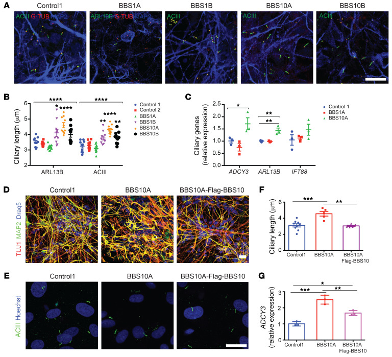
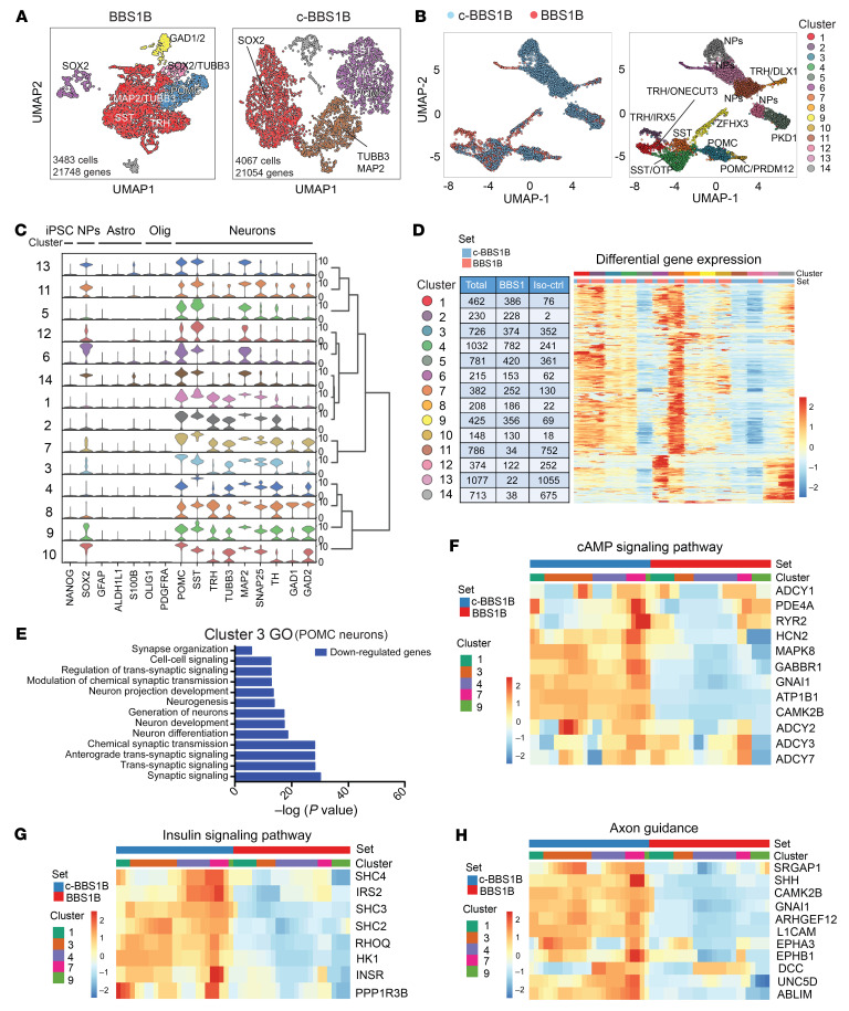
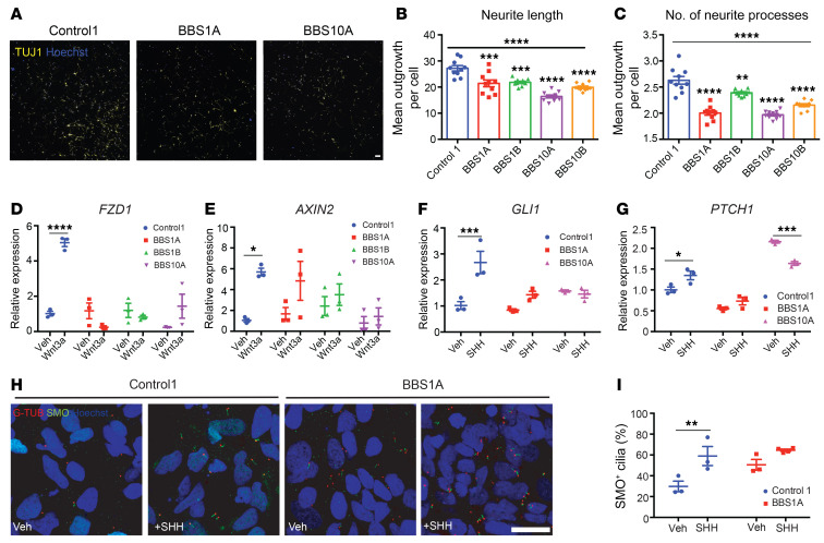
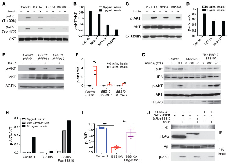
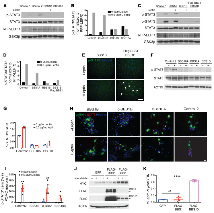
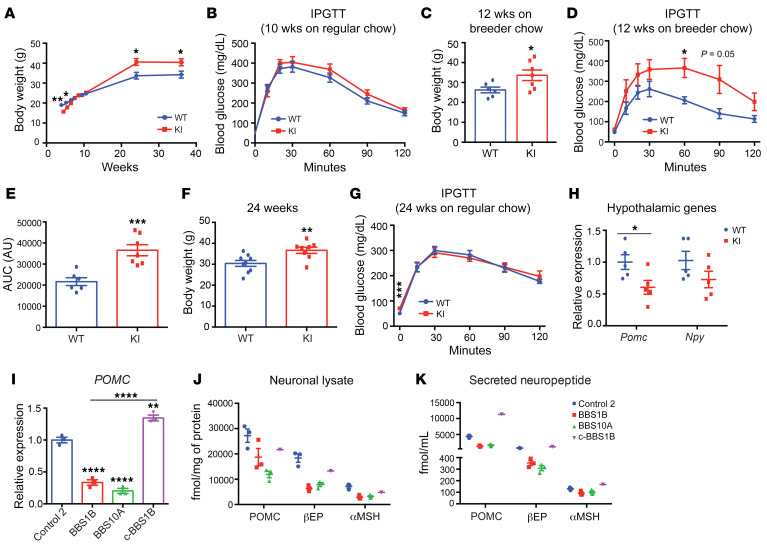
Bardet-Biedl syndrome (BBS) is a rare autosomal recessive disorder caused by mutations in genes encoding components of the primary cilium and is characterized by hyperphagic obesity. To investigate the molecular basis of obesity in human BBS, we developed a cellular model of BBS using induced pluripotent stem cell-derived (iPSC-derived) hypothalamic arcuate-like neurons. BBS mutations BBS1M390R and BBS10C91fsX95 did not affect neuronal differentiation efficiency but caused morphological defects, including impaired neurite outgrowth and longer primary cilia. Single-cell RNA sequencing of BBS1M390R hypothalamic neurons identified several downregulated pathways, including insulin and cAMP signaling and axon guidance. Additional studies demonstrated that BBS1M390R and BBS10C91fsX95 mutations impaired insulin signaling in both human fibroblasts and iPSC-derived neurons. Overexpression of intact BBS10 fully restored insulin signaling by restoring insulin receptor tyrosine phosphorylation in BBS10C91fsX95 neurons. Moreover, mutations in BBS1 and BBS10 impaired leptin-mediated p-STAT3 activation in iPSC-derived hypothalamic neurons. Correction of the BBS mutation by CRISPR rescued leptin signaling. POMC expression and neuropeptide production were decreased in BBS1M390R and BBS10C91fsX95 iPSC-derived hypothalamic neurons. In the aggregate, these data provide insights into the anatomic and functional mechanisms by which components of the BBSome in CNS primary cilia mediate effects on energy homeostasis.

Keywords: Obesity; Stem cells.


Conflict of interest statement

Conflict of interest: The authors have declared that no conflict of interest exists.

Figures

Figure 1
Figure 1. BBS mutations increase ciliary length in iPSC-derived neurons.
(A) Immunocytochemistry (ICC) staining of primary cilia in iPSC-derived neurons. Neurons were stained for MAP2 as well as ciliary markers: adenylate cyclase III (ACIII, basal body and axoneme), ARL13B (axoneme), and γ-tubulin (G-TUB, basal body). Scale bar: 20 μm. (B) Quantification of ciliary length in control and BBS iPSC–derived neurons in A (n = 10 for each line). *P < 0.05, **P < 0.01, ****P < 0.0001 by 1-way ANOVA followed by Bonferroni’s multiple-comparison test (compared with control 1). (C) Expression of ciliary genes in control and BBS iPSC–derived neurons (n = 3). *P < 0.05, **P < 0.01 by 1-way ANOVA followed by Tukey’s multiple-comparison test. (D) ICC staining of control 1, BBS10A, and BBS10A-FLAG-BBS10 iPSC–derived neurons. TUJ1 and MAP2 are neuronal markers. Scale bar: 20 μm. (E) ICC staining of primary cilia in FACS-isolated NCAM+ neurons. Cells were stained with anti-ACIII and Hoechst. Scale bar: 10 μm. (F) Quantification of ciliary length in E (n = 9, 5, and 6, left to right). (G) Ectopic expression of FLAG-BBS10 partially reverses ADCY3 expression in BBS10A iPSC–derived neurons (n = 3). *P < 0.05; **P < 0.01; ***P < 0.001 by 1-way ANOVA followed by Tukey’s multiple-comparison test (F and G).
Figure 2
Figure 2. scRNA-seq analysis of BBS iPSC–derived hypothalamic neurons.
(A) Uniform manifold approximation and projection (UMAP) of BBS1B and isogenic control (c-BBS1B) iPSC–derived hypothalamic neurons. (B) UMAP of BBS1B and c-BBS1B neurons after integration and identification of 14 clusters. Marker genes and cell types for each cluster are indicated. (C) Violin plots of cell-type-specific markers for all 14 clusters and hierarchical clustering. Signature genes for iPSCs, neuronal progenitors (NPs), astrocytes (Astro), oligodendrocytes (Olig), and neurons are included. (D) Heatmap of differentially expressed genes in all clusters. Number of cells from which line within each cluster is indicated in the table. (E) Gene Ontology analysis of downregulated genes (BBS1B/c-BBS1B) identified in cluster 3. (FH) Heatmaps of genes of pathways highlighted in KEGG pathway analysis. Genes involved in cAMP signaling pathway (F), insulin signaling pathway (G), and axon guidance (H) were plotted against cell set and cluster ID.
Figure 3
Figure 3. BBS mutations disturb neurite outgrowth and impair Wnt and SHH signaling in TUJ1+ iPSC–derived neurons.
(AC) Neurite outgrowth assay of control and BBS iPSC–derived neurons on day 30 of differentiation. (A) TUJ1 staining of control and BBS mutant cultures. Scale bar: 50 μm. Mean neurite length (B) and average number of neurite processes (C) were calculated using the neurite outgrowth tool in MetaMorph software based on TUJ1 and Hoechst staining (n = 10 independent wells, 2,500 cells/well). **P < 0.01, ***P < 0.001, ****P < 0.0001 by 1-way ANOVA followed by Bonferroni’s multiple-comparison test (vs. control 1); the asterisks above the horizontal bars in B and C are for the 1-way ANOVA that permitted the pair-wise testing. (D and E) Wnt signaling is impaired in BBS iPSC–derived neurons. Control 1, BBS1A, BBS1B, and BBS10A iPSC–derived neurons (day 30) were treated with vehicle (Veh) or 100 ng/mL Wnt3a for 16 hours. Frizzled 1 (FZD1) and AXIN2 mRNA levels were determined by qPCR (n = 3). (F and G) SHH signaling is reduced in BBS iPSC–derived neurons. Control and BBS iPSC–derived neurons (day 30) were treated with vehicle or 100 ng/mL SHH for 16 hours. GLI1 and PTCH1 mRNA levels were analyzed by qPCR (n = 3). (H) Smoothened (SMO) staining in SHH-treated control and BBS iPSC–derived neurons. Control and BBS1A iPSC–derived neurons were treated with vehicle or 100 ng/mL SHH overnight. Neurons were fixed and stained with anti-SMO and anti–γ-tubulin (G-TUB, basal body) antibodies for cilia and Hoechst for nuclei. Scale bar: 20μm. (I) Quantification of SMO+ cilia in E. Hoechst was used as nuclear marker. SMO+ cilia percentage was calculated by (SMO+ cells/Hoechst+ cells) × 100 (n = 3 independent images). *P < 0.05, **P < 0.01, ***P < 0.001, ****P < 0.0001 by 2-way ANOVA followed by Bonferroni’s multiple-comparison test (DG and I).
Figure 4
Figure 4. BBS1 and BBS10 bind to the insulin receptor and influence insulin signaling.
(A) BBS mutations disrupt insulin signaling in human fibroblasts. Western blot (WB) analysis of insulin signaling as indicated by phosphorylation of AKT in control and BBS fibroblasts. Fibroblasts were serum starved overnight and treated with 0 or 1 μg/mL insulin for 30 minutes. AKT, p-AKT Thr308, and p-AKT Ser473 were probed. (B) Quantification of p-AKT (Thr308)/AKT from A (n = 1). (C) BBS mutations abrogate insulin signaling in iPSC-derived TUJ1+ neurons. Control, BBS1A, and BBS10A iPSC–derived neurons (day 30) were serum starved overnight and treated with 0 or 1 μg/mL insulin for 30 minutes. p-AKT Ser473, AKT, and α-tubulin (A-TUB) were probed. (D) Quantification of p-AKT/AKT from WB in C (n = 1). (E) Diminished insulin signaling in neurons derived from 2 BBS10 shRNA-knocked-down iPSC lines as indicated. iPSC-derived TUJ1+ neurons (day 30) were serum starved overnight and treated with 0 or 1 μg/mL insulin for 30 minutes. p-AKT Ser473, AKT, and actin were examined by WB. (F) Quantification of p-AKT/AKT from WB in E (n = 2–3). (G) BBS10 disrupts insulin signaling by disturbing phosphorylation of the insulin receptor (IR) in day 12 neuronal progenitors (NPs). WB analysis of insulin signaling molecules as indicated in control, BBS10A, and BBS10A-FLAG-BBS10 transgenic NPs after 30-minute treatment with 0.01 and 0.1 μg/mL insulin. FLAG was used to confirm the overexpression of WT BBS10. (H and I) Quantification of p-AKT/AKT (H) (n = 1) and p-IR/IR (I) (n = 3) in D. **P < 0.01 by 1-way ANOVA followed by Tukey’s multiple-comparison test. (J) Coimmunoprecipitation (IP) confirms the interaction between BBS proteins and the IR. 293FT cells were transfected with CD615-GFP or CD615-3×FLAG-BBS1-GFP or CD615-3×FLAG-BBS10-GFP for 48 hours before insulin (1 μg/mL) treatment (30 minutes). IRβ, p-AKT Ser473, and FLAG were probed in FLAG IP samples and total input.
Figure 5
Figure 5. BBS mutations impair leptin signaling in RFP-LEPR transgenic human fibroblasts and iPSC-derived hypothalamic neurons.
(AD) Leptin signaling is impaired in RFP-LEPR transgenic BBS fibroblasts. Control 1, control 2, BBS1A, BBS1B, BBS10A, and BBS1B + FLAG-BBS1 RFP-LEPR transgenic fibroblasts were treated with 0 or 0.5 μg/mL leptin (30 minutes) after overnight serum starvation. Leptin signaling was assessed by Western blot (WB) of p-STAT3 levels. STAT3, RFP, and GSK3β were also probed (A and C). In C, the 2 p-STAT3 blot images are from short (top) and long time exposures. (B and D) Quantification of p-STAT3/STAT3 from A and C. p-STAT3/STAT3 was further normalized to RFP-LEPR/GSK3β (n = 1). (E) Leptin signaling can be rescued in BBS1B RFP-LEPR transgenic fibroblasts by overexpressing FLAG-BBS1. BBS1B and BBS1B + FLAG-BBS1 RFP-LEPR transgenic fibroblasts were treated as in C and stained with anti–p-STAT3 and Hoechst. Arrows indicate p-STAT3–positive cells. Scale bar: 200 μm. (F and G) Leptin signaling is disturbed in BBS iPSC–derived hypothalamic neurons. Day 34 iPSC-derived hypothalamic neurons were serum starved overnight and treated with 0 or 1 μg/mL leptin for 30 minutes. (F) p-STAT3, STAT3, and actin were examined by WB. (G) Quantification of WB in F (n = 2–3). (H and I) Leptin signaling, as measured by immunostaining of p-STAT3, is disrupted in BBS iPSC–derived hypothalamic neurons. (H) Day 34 neurons were treated with leptin and stained with anti-αMSH, anti–p-STAT3, and Hoechst. Scale bar: 20 μm. (I) Quantification of p-STAT3+ POMC neurons in H (n = 3). **P < 0.01 by 2-way ANOVA followed by Bonferroni’s multiple-comparison test. (J) Overexpression of BBS10 increases total LEPR proteins. 293FT cells were cotransfected with mouse LEPRB-Myc (mLeprb-Myc) and GFP or FLAG-BBS1 or FLAG-BBS10 for 24 hours. Myc, FLAG, and actin were probed. Arrows indicate FLAG-BBS1 and FLAG-BBS10 bands. (K) Quantification of mLeprb-Myc in J. ****P < 0.0001 by 1-way ANOVA followed by Tukey’s multiple-comparison test. NS, no significant difference.
Figure 6
Figure 6. BBS1M390R mutation reduces POMC expression in both mouse hypothalamus and human iPSC–derived hypothalamic neurons.
(A) Body weight curve of male WT and BBS1M390R-knockin (KI) mice (n = 9, 10). (B) Intraperitoneal glucose tolerance test (IPGTT) of 10-week-old male mice on regular chow diet (n = 9, 10). (C and D) Body weight (C) and IPGTT (D) of 12-week-old male mice fed breeder chow ad libitum (n = 6, 7). (E) The glucose area under the curve (AUC) in WT and KI mice as shown in D. ***P < 0.001. (F) Body weight of 24-week-old WT and KI mice on regular chow diet (n = 9, 8). (G) IPGTT of 24-week-old WT and KI mice on regular chow diet (n = 9, 8). (H) qPCR analysis of Pomc and Npy expression in hypothalamus of WT and KI mice (24-week-old males) after 16-hour fasting followed by 4-hour refeeding (n = 5, 5). *P < 0.05, **P < 0.01 by 2-tailed Student’s t test (AH). (I) qPCR analysis of POMC expression in day 35 iPSC-derived hypothalamic neurons (n = 3). **P < 0.01, ****P < 0.0001 by 1-way ANOVA followed by Tukey’s multiple-comparison test. (J and K) Amount of neuropeptide produced in neuronal lysates (J) and in cultured medium (16-hour culture, K) from control (n = 3), BBS1B (n = 3), BBS10A (n = 3), and c-BBS1B (n = 1) iPSC–derived hypothalamic neurons. POMC, αMSH, and βEP concentrations were measured with ELISA and radioimmunoassay and normalized to total protein.

Comment in

Similar articles

Cited by

References

    1. Tsang SH, et al. Ciliopathy: Bardet-Biedl syndrome. Adv Exp Med Biol. 2018;1085:171–174. doi: 10.1007/978-3-319-95046-4_33. - DOI - PubMed
    1. M’Hamdi O, et al. Update on the genetics of Bardet-Biedl syndrome. Mol Syndromol. 2014;5(2):51–56. doi: 10.1159/000357054. - DOI - PMC - PubMed
    1. Jin H, et al. The conserved Bardet-Biedl syndrome proteins assemble a coat that traffics membrane proteins to cilia. Cell. 2010;141(7):1208–1219. doi: 10.1016/j.cell.2010.05.015. - DOI - PMC - PubMed
    1. Wingfield JL, et al. Trafficking of ciliary membrane proteins by the intraflagellar transport/BBSome machinery. Essays Biochem. 2018;62(6):753–763. doi: 10.1042/EBC20180030. - DOI - PMC - PubMed
    1. Liu P, Lechtreck KF. The Bardet-Biedl syndrome protein complex is an adapter expanding the cargo range of intraflagellar transport trains for ciliary export. Proc Natl Acad Sci U S A. 2018;115(5):E934–E943. doi: 10.1073/pnas.1713226115. - DOI - PMC - PubMed

Publication types

MeSH terms